Download

1 / 57
570 likes | 593 Vues
Learn about the principle and performance of electrophoresis techniques for nucleic acids, including agarose and polyacrylamide gel electrophoresis, capillary electrophoresis, and pulsed field gel electrophoresis. Discover the various detection systems used in nucleic acid applications.

E N D
Chapter 5: Resolution and Detection of Nucleic Acids Donna C. Sullivan, PhD Division of Infectious Diseases University of Mississippi Medical Center
Objectives • Explain the principle and performance of electrophoresis as it applies to nucleic acids. • Compare and contrast agarose and polyacrylamide gel polymers. • Explain the principle and performance of capillary electrophoresis as it is applies to nucleic acid separation. • Describe the general types of equipment used for electrophoresis. • Discuss methods and applications of pulsed field gel electrophoresis. • Compare and contrast detection systems used in nucleic acid applications.
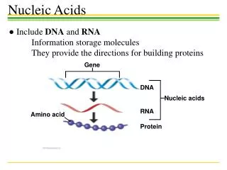
Gel Electrophoresis • Electrophoresis is the movement of molecules by an electric current. • Nucleic acid moves from a negative to a positive pole. • Nucleic acid has a net negative charge, they RUN TO RED
Electrophoresis of Nucleic Acids • Nucleic acids are separated based on size and charge. • DNA molecules migrate in an electrical field at a rate that is inversely proportional to the log10 of molecular size (number of base pairs). • Employs a sieve-like matrix (agarose or polyacrylamide) and an electrical field. • DNA possesses a net negative charge and migrates towards the positively charged anode.
Applications of Electrophoretic Techniques in the Molecular Diagnostics Laboratory • Sizing of Nucleic Acid Molecules • DNA fragments for Southern transfer analysis • RNA molecules for Northern transfer analysis • Analytical separation of PCR products • Detection of Mutations or Sequence Variations
Principles of Gel Electrophoresis • Electrophoresis is a technique used to separate and sometimes purify macromolecules • Proteins and nucleic acids that differ in size, charge or conformation • Charged molecules placed in an electric field migrate toward either the positive (anode) or negative (cathode) pole according to their charge • Proteins and nucleic acids are electrophoresed within a matrix or "gel"
ELECTROPHORESIS DNA and RNA are negatively charged; they RUN TO RED!
Principles of Gel Electrophoresis • The gel itself is composed of either agarose or polyacrylamide. • Agarose is a polysaccharide extracted from seaweed. • Polyacrylamide is a cross-linked polymer of acrylamide. • Acrylamide is a potent neurotoxin and should be handled with care!
Agarose Acrylamide Gel Electrophoresis Matrices
Types Of Nucleic Acid Electrophoresis • Agarose gel electrophoresis • DNA or RNA separation • TAE or TBE buffers for DNA, MOPS with formaldehyde for RNA • Polyacrylamide gel electrophoresis (PAGE) • Non-denaturing (Special applications in research) • Denaturing contain 6-7 M Urea (Most common)
Agarose Gel Electrophoresis • Separates fragments based on mass, charge • Agarose acts as a sieve • Typically resolve 200 bp-20 kbp • fragments <200 bp, polyacrylamide gels • fragments> 20 kbp, pulse field gels • Include DNA size standards
Factors That Effect Mobility Of DNA Fragments In Agarose Gels • Agarose Concentration • Higher concentrations of agarose facilitate separation of small DNAs, while low agarose concentrations allow resolution of larger DNAs (Remember-inversely proportional!) • Voltage • As the voltage applied to a gel is increased, larger fragments migrate proportionally faster that small fragments • Charge is evenly spread (uniform) so the larger fragments will have more charged groups
Factors That Effect Mobility Of DNA Fragments In Agarose Gels • Electrophoresis Buffer • The most commonly used for double stranded (duplex) DNA are TAE (Tris-acetate-EDTA) and TBE (Tris-borate-EDTA). • Effects of Ethidium Bromide • Staining dye that inserts (intercalates) into the DNA between the nitrogenous bases (“rungs of the ladder”) and glows when exposed to UV light • Binding of ethidium bromide to DNA alters its mass and rigidity, and therefore its mobility
% agarose: 2% 4% 5% 500 bp 200 bp 50 bp 500 bp 200 bp 50 bp 500 bp 200 bp 50 bp Comparison of Agarose Concentrations
Gel Electrophoresis: The Basics • The movement of molecules is impeded in the gel so that molecules will collect or form a band according to their speed of migration. • The concentration of gel/buffer will affect the resolution of fragments of different size ranges. • Genomic DNAs usually run as a “smear” due to the large number of fragments with only small differences in mass
A B Agarose Electrophoresis of Restriction Enzyme Digested Genomic DNA
Gel Electrophoresis: Apparatus and Types of Gels • Horizontal Gel Units (“Submarine Gels”) • Most DNA and RNA gels • Agarose • Vertical Gel Units • Polyacrylamide gels • Typically sequencing gels • Pulse Field Gel Units • Any electrophoresis process that uses more than one alternating electric field • Agarose • Large genomic DNA (Chromosomal)
DNA/RNA is negatively charged: RUN TO RED Electrophoresis Equipment: Horizontal or Submarine Gel
DNA/RNA is negatively charged: RUN TO RED Agarose Gel Electrophoresis
Reservoir/Tank Power Supply Casting Tray and Combs www.biorad.com Agarose Gel ElectrophoresisHorizontal Gel Format
Reservoir/Tank Power Supply Glass Plates, Spacers, and Combs www.biorad.com Vertical Gel Format: Polyacrylamide Gel Electrophoresis
Electrophoresis Equipment • Combs are used to put wells in the cast gel for sample loading. • Regular comb: wells separated by an “ear” of gel • Houndstooth comb: wells immediately adjacent
Field inversion gel Transverse alternating field Crossed field (Reverse) Contour-clamped homogeneous electric field Types Of Pulse Field Gel Electrophoresis
Pulse Field Gel Electrophoresis • Used to resolve DNA molecules larger than 25 kbp • Periodically change the direction of the electric field • Several types of pulsed field gel protocols • FIGE: Field inversion gel electrophoresis • TAFE: Transverse alternating field electrophoresis • RGE: Crossed field electrophoresis • CHEF: Contour-clamped homogeneous electric field
Critical Parameters: Pulse Field Gel Electrophoresis • Depend on time it takes molecules of various sizes to change directions in a gel • Small DNA molecules are sieved (pass through the pores in the agarose gel) • Large DNA molecules are not “sieved” but “squeezed” through the gel at about the same rate, called the limiting mobility
Size of Fragments and Distance Traveled Not Linear When Large Fragments Are Analyzed
Pulse Field Gel Electrophoresis • PFGE works by periodically altering the electric field orientation • The large extended coil DNA fragments are forced to change orientation • Size dependent separation is re-established because the time taken for the DNA to reorient is size dependent
- - - - + + + + Comparison of Migration: Horizontal vs. CHEF
Preparation Of Intact DNA For PFGE • Conventional techniques for DNA purification (organic extraction, ethanol precipitation) produce shear forces • DNA purified is rarely greater than a few hundred kb in size • This is clearly unsuitable for PFGE which can resolve mb DNA • The problem of shear forces was solved by performing DNA purification from whole cells entirely within a low melting temperature (LMT) agarose matrix
Preparation Of Intact DNA For PFGE • Intact cells are mixed with molten low melting point (LMT) agarose and set in a mold forming agarose ‘plugs’ • Enzymes and detergents diffuse into the plugs and lyse cells • Proteinase K diffuses into plugs and digests proteins • If necessary restriction digests are performed in plugs (extensive washing or PMSF treatment is required to remove proteinase K activity) • Plugs are loaded directly onto PFGE and run
CHEF: Contour-Clamped Homogenous Gel Electrophoresis • Based on hexagonal array of alternate electric fields at 120 degree angle • Generates a more uniform electric field when compared to other PFGE systems • Programmable, autonomously controlled electrodes • Extremely versatile system based on CHEF hexagonal array • All electrophoretic parameters can be controlled at each electrode • Can generate electric field and switching characteristics of any PFGE system
Using PFGE In The Molecular Investigation Of An Outbreak Of S. marcescens Infection In An ICU • An outbreak due to S. marcescens infection was detected in the ICU • A total of 25 isolates were included in this study: • 12 isolates from infected patients • nine isolates from insulin solution • one isolate from sedative solution • one isolate from frusemide solution • two isolates from other wards which were epidemiologically-unrelated Singapore Med J 2004 Vol 45(5) : 214
Using PFGE in the Molecular Investigation Of An Outbreak of S. marcescens Infection in an ICU Singapore Med J 2004 Vol 45(5) : 214
Using PFGE in the molecular investigation of an outbreak of S. marcescens infection in an ICU • The S. marcescens from patients, insulin solution and sedative solution showed an identical PFGE fingerprint pattern. • The isolate from the frusemide solution had a closely-related PFGE pattern to the outbreak strain with one band difference. • Found that the insulin and sedative solutions used by the patients were contaminated with S. marcescens and the source of the outbreak. Singapore Med J 2004 Vol 45(5) : 214
Comparison Of Agarose Gel And PFGE Panel B: Agarose gel electrophoresis Panel C: PFG electrophoresis Pulsed Field Gel Electrophoresis was applied to the study of Duchenne Muscular Dystrophy. Since the DMD gene is 2.3Mbp, it was necessary to use PFGE in order to uncover the genetic defect. The use of PFGE analysis on patients with the disease soon revealed that in 50% of the cases large deletions or duplications were a responsible for the disease (Mathew, 1991).
Polyacrylamide Gel Electrophoresis (PAGE) • PAGE is the preferred method for PROTEINS but can be used for DNA/RNA • Gel prepared immediately before use by copolymerization of acrylamide and N,N'-methylene bis acrylamide under UV light. • Porosity controlled by proportions of the two components. • Larger pore size for larger proteins. • Gradient gels also possible.
Electrophoresis of Nucleic Acids Polyacrylamide Gel Electrophoresis (PAGE) • Advantages • High degree of resolving power. • Can effectively and reproducibly separate molecules displaying 1 bp differences in molecular size. • Optimal separation is achieved with nucleic acids that are 5–500 bp in size.
Electrophoresis of Nucleic Acids Polyacrylamide Gel Electrophoresis (PAGE) • Typical Conditions • Vertical gel setup, TBE buffer (Tris-borate/EDTA) and constant power. • Disadvantages • Acrylamide monomer is a neurotoxin. • Polyacrylamide gels can be difficult to handle.
Electrophoresis of Nucleic AcidsAgarose Gel Electrophoresis • Advantages • Greater range of separation of nucleic acid molecules. • Optimal separation is achieved with nucleic acids that are 200 bp to 30 kb in size. • Ease of preparation and handling.
PAGE: Critical Parameters • Polymerization reaction critical • High grade acrylamide, bis-acrylamide • Break down into acrylic acid (long shelf life solutions incorporate inhibitors of polymerization) • Must have even heat distribution to prevent “smiling”
PAGE: DNA • High resolution of low molecular weight nucleic acids (500bp) • Polymerization of acrylamide monomers into long chains • Cross link chains with bis-acrylamide • Initiated by free radicals in ammonium persulfate, stabilized by TEMED • Pore size determined by % acrylamide