Download

1 / 51
1.37k likes | 6.82k Vues
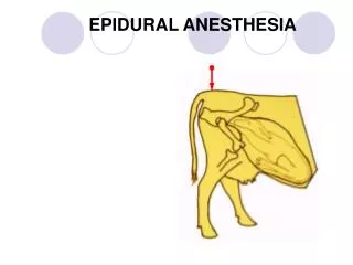
EPIDURAL TECHNIQUE. Definition. Epidural anesthesia is anesthesia obtained by blocking spinal nerves in the epidural space as the nerves emerge from the dura and then pass into the intervertebral foramina. The anaesthetic solution is deposited out side the dura. HISTORY:.

E N D
Definition • Epidural anesthesia is anesthesia obtained by blocking spinal nerves in the epidural space as the nerves emerge from the dura and then pass into the intervertebral foramina. • The anaesthetic solution is deposited out side the dura.
HISTORY: • First approach to epidural space was tried by cathelin & Tuffier (1901). • It was refined by Fidel Pages. • In 1921, Dr. Jacques Forestier and Sicard identified the space by attaching a syringe of fluid to the needle advancing through the ligaments. • Dogliotti in 1933 popularized the above test as Loss of Resistance’ test. • Guieterrez and Soresi (1933) were the first to apply internal pressure differences to identify the space and they devised ‘Hanging drop’ sign.
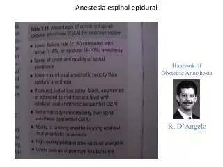
Contraindications: Epidural Blockade Absolute • Patient refusal • Uncorrected hypovolemia • Increased ICP • Infection at site • Allergic to LA Relative • Coagulopathy • Platelet count <1,00,000 • Uncooperative patient • Severe anatomic abnormalities of spine • Sepsis • Hypertension
Technical procedure • Preparation and monitoring of patient: -Functional IV line n emergency drugs -Blood pressure monitoring. -Equipment for airway management and O2 administration.
Equipment Solution of antiseptic. Syringe/needle for skin localization. Epidural needle. Glass syringe. Epidural catheter. Glass filter. Dosing syringe. Local anesthetic. Saline.
Ralph Huber • Dentist
Epidural needles • A styletted Tuohy epidural needle is typically 16 to 18 gauge, 8 cm in length, with surface markings at 1-cm intervals. • It has a shaft,a small hub with luerlok connector. • It has a 15 to 30degree curve at the tip with a blunt bevel. The curved Huber tip is designed to prevent accidental dural puncture and to control the direction that the cathetermoves in the epidural space. A: Hustead 17-gauge, B: Hustead 18-gauge, C: Crawford, D: Tuohy.
At the junction of the needle shaft and hub are wings to allow better control as the needle is passed through tissue. • Longer needles up to 10 cm in length are available for obese patients.
Hustead needle • It’s a modified Tuohyneedlewith a rounded tip and a bevel opening,the edges of which are smoother to reduce the incidence of catheter shearing.
Crawford needle- • tip is straight ,has a short bevel with smooth edges. • made of stainless steel with 2% molybdenum which improves resistance to corrosion.suitable for single shot tech.
Other needles such as CHENG AND CRAWLEY WANGER are less commonly used winged needles are ideal for hanging drop method
Epidural catheters • Available in different sizes 16G 18G 19G 20G • Material : Nylon,polyurethane,polyethylene and Teflon • Single lumen(open end) • Multi orifice(blunt tip)
Bromage ideal characteristics of an epidural catheter: • Biochemical inertness • Low coefficient of friction • High tensile strength • Maneuverable rigidity • Kink resistant • Atraumatic tip • Depth indicators • Radio opacity
Patient Positioning • Sitting PositionIf the sitting position is chosen, the patient should be assisted to sit on the edge of the table or bed with feet resting on a stool . • The patient should lean forward with elbows resting on a pillow or on the thighs. The back should be maximally flexed to open up the lumbar vertebral spaces. Flexing the neck will help the patient to flex the lower spine. • The assistant should help the patient to hold this position during the entire procedure.
Lateral Decubitus Position • In the lateral decubitus position, the patient is placed on her side with the back at the edge of the operating table that is closest to the anesthesiologist. • The spinous processes should be oriented parallel to the floor to prevent rotation of the spine. • The thighs should be flexed on the abdomen with the knees drawn to the chest and the neck flexed so that the chin rests on the chest. • Asking the patient to “assume the fetal position” or “touch your knees with your chin” may help with positioning during lumbar epidural placement.
Site of injection • A spinal interspace is chosen one to two segments below the middle of area to be anesthetized.
PREPARATION • Skin is cleaned ,an antiseptic applied, and the area draped ,a skin wheal is made and deeper tissues infiltrated with 2% lidocaine
Approach • MIDLINE APPROACH This approach is most commonly used for lumbar or low thoracic epidural placement . • PARAMEDIAN APPROACH Patients who cannot be positioned easily or cannot flex the spine Calcified interspinous ligament Spine deformities: kyphoscoliosis, prior lumbar surgery
Insertion of needle High thoracic region. Low thoracic region Lumbar region.
Insertion of needle • Insert epidural needle with stylet through same skin puncture. The dorsum of the anesthesiologist’s non injecting hand rests on the patient’s back with the thumb and index finger holding the hub of the epidural needle (Bromage grip). • Advance the needle through the supraspinous ligament and into the interspinous ligament at which point the needle should sit firmly in the midline • After the ligaments are penetrated, it is no longer possible to change the direction of the needle tip
A glass syringe or a low resistance plastic syringe is filled with 2-3 ml saline /air and attached to hub of epidural needle ,after removing the stylet. • Maintain constant pressure on testing syringe with the dominant hand. • Controlled needle advancement is made with the non dominant hand. • As the needle enters the epidural space there is sudden loss of resistance as the saline or air is rapidly injected.
Detection of epidural space • Negative pressure techniques. • Loss of resistance technique. Negative pressure techniques 1 Hanging drop sign: a small drop of sterile water is placed on the hub of the needle with entry into epidural space, this drop will be sucked into the epidural space. this is called ‘sign of drop’
Capillary tube method: Odom devised a small capillary tube filled with sterile saline in which one or two bubbles of air were placed. as soon as needle enters epidural space, saline will be sucked in, and air bubbles could be seen to advance into the space
Manometer technique • a small U shaped tube about 3-4 inches high is used as a water manometer after needle has been introduced into the interspinous ligaments ,manometer is attached to needle. as it is advanced through lig flavum and enters the epidural space ,there is an immediate movement of the liquid ,signifying a negative pressure
Loss of resistance technique • Syringe technique sudden loss of resistance to a pressure exerted on plunger of a syringe filled with water as a needle advances through the ligamentum flavum was first recognized by Sicard and Forestier Liquids transmit pressure changes more quickly than air
Others technique are • Spring loaded syringe • Saline drip technique • Balloon technique • Brooks device (odoms indicator) • Vertical Tube of Dawkins
Confirmatory Test for Epidural Puncture 1. Aspiration Test: Gentle suction is done with 2m1 syringe. CSF or blood can be easily detected. 1-2 ml of air is injected through needle and aspiration is again performed. Air should go easily, but nothing should return on aspiration. 2. Sterile water injection: Fluids which differ from normal tonicity are painful in epidural space — (Lund’s concept).
3. Rapid injection of NS ( or) LA:- • Rapid injections when given epidurally in conscious patients cause an increase in CSF pressure leading to feelings of discomfort and anxiety. In unconscious patient, the rate and depth of respiration is increased.
Once a loss of resistance to air or saline has occurred, the glass syringe is removed, and depth at which the epidural space was entered is noted. The noninjecting hand should continue to hold the needle in place. • Note the depth of the needle at the skin. The marking on the needle at the skin is the depth from the skin to the epidural space.
Thread the catheter gently through the needle into the epidural space to approximately the 15- 17-cm mark, then remove the needle without dislodging the catheter • Add the skin-to-epidural depth plus 3–5 cm. Withdraw the catheter to that point and secure. No more than 5 cm of catheter should be left in the epidural space to prevent displacement of the catheter laterally or into extradural structures.
PARAMEDIAN APPROACH The needle entry site is marked approximately 1.5–2 cm lateral and caudal to the desired level of blockade Epidural needle angulation 45 degrees cephalad and very slightly medial. When (if) the bone (lamina) is contacted during needle advancement, the cephalad needle angle is lowered to walk off the lamina.
Midthoracic Epidural Paramedian Approach • In the midthoracic level, the skin wheal is placed 2.0 cm lateral and inferior to the superior spinous process • Epidural needle is advanced perpendicularly through the skin at the same location until the lamina is contacted. • The needle is withdrawn approximately 2 cm, redirected at a 15- to 20-degree angle toward the midline and a 45-degree angle from the skin surface. • Each time bone is contacted, the needle is withdrawn 0.5 cm, then walked off the bone in a medial/cephalad direction until the ligamentum flavum is entered
CAUDAL APPROACH • Patient is placed in a lateral or prone position (pillow under pelvis if prone). • Either a smaller gauge IV catheter (18- to 23-gauge) or a 20-gauge epidural needle is advanced at a 45-degree angle from the back with the bevel up (to avoid penetrating the anterior sacral wall). • A distinct “pop” or “snap” is felt when the needle pierces the sacrococcygeal membrane. • The needle angle is lowered to 160 degrees (almost flat) toward the back. It is advanced not more than 1.5 cm (usually between 5 and 7 mm) in adults and not more than 0.5 cm in children. • Aspirate for blood or CSF before injecting local anesthetic. • The epidural catheter can then be inserted through the needle to the desired level
Test dose • Should consist of 3ml of local anaesthetic, to which 15µg of epinephrine(5µg/ml) is added, • To reveal accidental subarachnoid/IV injection. • Subarachnoid injection is made out by motor blocked • CRITERIA FOR POSITIVE EPINEPHRINE TEST (ASRA 2001)
Drug dosage • 150cm 1ml/seg ,2% lignocaine/equipotent dose of other drugs +5cm-↑0.1ml/seg Dose is ↓ 30% in pregnancy &obese 20-40yr 1-1.5ml/seg adjusted to ht 40-60yr 0.5-1ml/seg rule of thumb 60-80yr 0.3-0.6ml/seg
Top up dose • Monitoring for signs of segmental regression will indicate the need for top up dose • Approximate time for regression of analgesia for top up dose lignocaine -60 min bupivacaine-120min • If initial dose is too high top up dose should be appropriately delayed
Complications Of Epidural Blockade Drug-related or Procedure-related. • When an excessive dose of local anesthetics is injected into the epidural space or when a moderate dose is accidentally injected into an epidural vein. • central nervous system is the first system affected lightheadedness, tinnitus, circumoral numbness and tingling, numbness of the tongue, and blurred vision. Signs -muscle twitching, confusion, tremors of the facial muscles and extremities, and shivering.
Cardiovascular effects of local anesthetics range from mild changes in blood pressure and pulse to complete cardiovascular collapse. • Treatment is supportive directed toward maintaining the airway, supporting ventilation, and cardiopulmonary resuscitation • prevent hypoxia, hypercarbia, and acidosis to limit the cardiovascular toxic effects of systemically administered local anesthetics.
Procedure-Related Complications • Subarachnoid Injection/High or Total Spinal • large dose of local anesthetic is given directly into the CSF • Profound hypotension, bradycardia, and apnea will occur. Unconsciousness follows as a result of the effect of local anesthetic action on the brainstem. • Treatment includes airway support and intubation, 100% oxygen, intravenous fluids and vasopressors to maintain hemodynamic stability
Neurologic Complications Associated with Epidural Anesthesia/Analgesia • Spinal nerve neuropathy • Anterior spinal artery syndrome • Adhesive arachnoiditis • Epidural hematoma • Epidural abscess
conclusion • Epidural placement is a safe, effective means of providing surgical anesthesia or postoperative analgesia. • It reduces the adverse physiologic responses to surgery, may decrease the incidence of myocardial infarctions and postoperative pulmonary sequelae. • Mastery of epidural placement comes with practice, attention to detail, and persistence. A thorough knowledge of anatomy, physiology, and the pharmacology of anesthetic agents is required for safe application.