Download
1 / 40
441 likes | 608 Vues
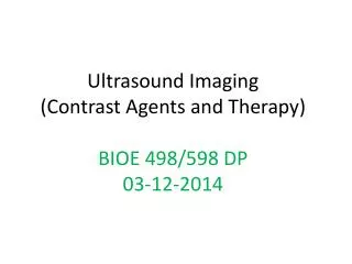
Ultrasound Imaging (Contrast Agents and Therapy) BIOE 498/598 DP 03-12-2014. Ideal Characteristics of an Ultrasound Probe. High echogenicity Low attenuation Low blood solubility Low diffusivity Ability to traverse pulmonary system Lack of biological effects in repeat exposures.

E N D
Ultrasound Imaging(Contrast Agents and Therapy)BIOE 498/598 DP03-12-2014
Ideal Characteristics of an Ultrasound Probe • High echogenicity • Low attenuation • Low blood solubility • Low diffusivity • Ability to traverse pulmonary system • Lack of biological effects in repeat exposures
Contrast Agents and Their Distribution Relative to the Vascular Endothelial Cells
Diagram illustrating development stage of microbubbles, nanobubbles, and nanodroplets for diagnostic and therapeutic purposes. HIFU = high-intensity focused ultrasound; KDR = kinase domain receptor.
Microbubbles Response to US Microbubblesvolumetrically oscillate due to continuous changes in the external pressure caused by an acoustic sound wave. At high pressure amplitudes, microbubbles collapse violently, producing water jetting and shock waves, and other inertial phenomena. Lindner JR. Microbubles in medical imaging: current applications and future directions. Nat Rev Drug Discov. 2004 Jun;3(6):527-32.
A slow motion capture of interial cavitations of micro bubbles Contrast harmonic image of liver in vivo Right frame:Image using both tissue and contrast agent fundamental frequency backscatter Left frame:the fundamental tissue signal has been filtered leaving the higher harmonic signal corresponding only to the contrast agent and, subsequently, the blood flow of the liver.
Acoustic Properties of Microbubbles Lindner JR. Microbubles in medical imaging: current applications and future directions. Nat Rev Drug Discov. 2004 Jun;3(6):527-32.
Ultrasonic Molecular Imaging of Tumor Angiogenesis in Breast Cancer Orthotopically implanted human breast adenocarcinomaxenograft (MDA-MB-231 cells) in nude mouse imaged following intravenous administration of 5×10⁷ VEGFR2-targeted probe. Scale bar =3 mm Bubbles coalescing in the extravascular space become detectable by US due to their micron sized collections
Albunex • Air-filled sonicated albumin MBs (MBI, San Diego, California) • 1994: First ultrasound CA approved for use in US • Albumin shell designed to prevent outward diffusion of air from MBs • Substantial loss of gas volume occurred during transit to systemic circulation following intravenous injection • Markedly decreased contrast enhancement and short duration of clinically useful contrast
Newer Agents • Newer agents designed to improve intravascular stability • Modifications in shell and gas content • “Air-tight” polymer shells or lipid-galactose stabilized shells designed to minimize outward diffusion of gas • Use of gases less prone to outward diffusion than air • Inert high-molecular-mass gases with low diffusion coefficients and low solubility in water (low Ostwald coefficient); result in prolonged lifespan of MBs
OptisonTM • Perflutren Protein-Type A Injectable Microspheres • GE Healthcare, Buckinhamshire, UK • Octafluoropropane • Manufacturer has voluntarily suspended marketing since 2005 Structural Formula Optison with RBCs http://www.amershamhealth-us.com/optison/monograph/om03-02.html
Definity • FDA approval in 2001 • Bristol-Myers Squibb Medical Imaging, Billerica, MA • $65 million in sales in 2006 • More than 2 million patients dosed
Subcutaneous human ovarian adenocarcinoma xenograft tumor (arrows) imaged with targeted contrast-enhanced ultrasound imaging after intravenous injection of integrin-targeted (A) and MB- (control) (B). Imaging signal obtained after MB-targeted injection was substantially higher than that obtained after control injection.
MBs Loaded with Chemotherapeutics Docetaxel The prepared nanoparticles in vitro released the drug steadily for 72 hours without occurrence of burst release.
Influence of ultrasound exposure on tumor temperature After ultrasound exposure, the tumor temperature increased steadily in a time dependent manner. The growth curve of hepatocellular carcinoma xenografts.The tumor volume increased persistently in model group, while in all the docetaxel treated groups, the tumor volume increment was inhibited, especially in the GDN+UE group.
Laplace Pressure The Laplace pressure is the pressure difference between the inside and the outside of a curved surface. The pressure difference is caused by the surface tension of the interface between liquid and gas. The Laplace pressure is determined from the Young–Laplace equation given as where R1 and R2 are the radii of curvature and is the surface tension.
Phase-change contrast agents (PCCAs) Consist of perfluorocarbons (PFCs) that are initially in liquid form, but can then be vaporized with acoustic energy. • Exposing pre-formed PFC micro bubbles to decreased ambient temperature and increased ambient pressure results in condensation of the gaseous core. • The decreased size results in an increased Laplace pressure, which serves to preserve the particle in the
Phase-change contrast agents (PCCAs) Exposing pre-formed PFC micro bubbles to decreased ambient temperature and increased ambient pressure results in condensation of the gaseous core. The decreased size results in an increased Laplace pressure, which serves to preserve the particle in the PCCAs take advantage of the volumetric increase that occurs during vaporization. Microscaledroplets limited to flow in the vascular space have been proposed for a number of techniques, including temporary occlusion of blood vessels and enhancing cavitationactivity. PCCAs manufactured with diameters in the 100-nm range may be able to diffuse out by EPR before phase-transition Contrast-enhanced extravascular imaging, cell-specific targeting, drug delivery, and therapy via ultrasound.
A microscale droplet of MBs exposed to a 0.25 µs pulse at approximately 0.55 MPa vaporizes to form a gas bubble approximately 5-fold larger
Bright field (a, c) and fluorescence (b, d) microscopy illustrating that the shell is preserved when DiI-labeled DFB microbubbles (a, b) are condensed to the liquid state (c, d), the shell is preserved through the change in volume.
Therapy with PCCA Schematic illustration of drug transfer from nanodroplets through microbubbles into cells under the action of ultrasound. PFP/PEG-PLLA microbubbles in a plasma clot before (A) and after sonication for 1 min by 1-MHz, 3.4 W/cm2 (B) and 90-kHz, 2.8 W/cm2 ultrasound (C) at room temperature.
Fig. 2A. PEG-PCL nanodroplets inserted in PBS through a 18 G needle (left) or 26 G needle (right). Bubbles formed when nanoemulsion is injected through a thin needle are visualized as bright spots (indicated by arrows in the right panel); bubbles rise to the sample surface while droplets precipitate to the bottom of a test tube. Fig. 2B. PEG-PCL nanodroplets injected in the agarose gel through a 18 G (left) or 26 G (right) needle. Injection through the thin needle results in immediate formation of very bright bubbles whose size and brightness increase with time; the brightness of the droplets also increases with time suggesting a post-injection droplet-to-bubble transition.
Photographs of a mouse bearing two ovarian carcinoma tumors (A) - immediately before and (B) - three weeks after the treatment; a mouse was treated by four systemic injections of nanodroplet-encapsulated PTX, nbGEN (20 mg/kg as PTX) given twice weekly; the right tumor was sonicated by 1-MHz CW ultrasound (nominal output power density 3.4 W/cm2, exposure duration 1 min) delivered 4 hours after the injection of the drug formulation. Ultrasound was delivered through a water bag coupled to a transducer and mouse skin by Aquasonic coupling gel.
Effective regression of the ovarian carcinoma tumor treated by nbGEN/ultrasound combination as described in Materials and Methods. The first photograph was taken before the start of the treatment, the second – two weeks later, i.e. immediately after the last treatment of the first treatment round. The third photograph was taken one week after the completion of the first treatment round. Fig. 6B. Normalized tumor growth/regression curve for the mouse presented in Figure 6A.
Ultrasound and pH Dually Responsive Polymer Vesicles for Anticancer Drug Delivery Upon ultrasound radiation, the disruption and re-self-assembly lead to smaller vesicle. Upon decreasing the solution pH, the shrinkage of vesicles by the recrystallization of PTMA chains surpasses the swelling of vesicles by the partial protonation of DEA, leading to smaller vesicles. Further decreasing the solution pH will lead to the complete protonation of DEA and finally the disassembly of vesicles. Both ultrasound radiation and decreasing pH can lead to faster drug release.
A) Recanalization was observed in angiograms taken post-HIFU treatment. 2/4 (50%) of animals treated with 415 W power, exhibited recanalization when compared to post-stroke angiograms. There was no damage or leakage from the vessel apparent on the angiogram. B) Post-treatment H&E showed no evidence of tissue damage or hemorrhage due to HIFU in the region of sonication or in the surrounding brain tissue. Scale bar = 500 µm. C) Prussian blue staining was used to detect small fragments of the disrupted iron-loaded blood clots. doi:10.1371/journal.pone.0042311.g003
Intravital fluorescence images of subcutaneous pancreatic tumors before and after FUS treatment
(A) and (B) – two coronal slices in the 19F MR images superimposed on the low resolution proton anatomic images of a pancreatic tumor bearing mouse. A 2% PFCE nanoemulsion stabilized with PEG-PCL copolymer was systemically injected every two hours, 200 μl each. Four injections were given, for a total of the PFCE dose of 2 mmol/kg. Images were recorded an hour after the last nanodroplet injection (seven hours after the start). (C)- Multiple liver metastases (arrows) are revealed at the necropsy of the mouse (indicated by long thing arrows). Transparent large tumor is indicated by a thick arrow. Organs could be displaced at necropsy.
Dramatic regression of a breast cancer MDA MB231 tumor treated by four systemic injections of paclitaxel loaded 1% PFCE/0.25% PEG-PCL nanoemulsion and focused 1-MHz CW ultrasound applied for 60 s
FDA “Black Box” Warning • Issued on October 10, 2007 • Post-marketing reports of 11 deaths 1-12 hours following administration of perflutren-based contrast agents • 10 patient deaths following Definity injection and 1 death following Optison injection • 4 patient deaths temporally related to contrast injection • Perflutren-based compounds contraindicated for use in patients with: 1. Acute coronary syndromes 2. Acute myocardial infarction 3. Worsening or clinically unstable heart failure http://www.fda.gov/cder/drug/InfoSheets/HCP/microbubbleHCP.htm
Safety Data Kitzman DW, Goldman ME, Gilliam LD, Cohen JL, Aurigemma GP, Gottdiener JS. Efficacy and safety of the novel ultrasound contrast agent perflutren (definity) in patients with suboptimal baseline left ventricular echocardiographic images. Am J Cardiol. 2000 Sep 15;86(6):669-74.
Safety Data • No clinically significant change in physical examination, vital signs, electrocardiographic tracings, or chemistry or hematology laboratory values • Adverse event rates similar across treatment groups • 30 of 169 patients (18%) in the combined perflutren-treated group (15% in the 5 ml/kg group and 20% in the 10 ml/kg group) • 6 of 42 placebo-treated patients (14%) • Headache was most frequently reported adverse event (9 of 169 patients who received perflutren (5%) and 3 of 42 patients who received placebo (7%) Kitzman DW, Goldman ME, Gilliam LD, Cohen JL, Aurigemma GP, Gottdiener JS. Efficacy and safety of the novel ultrasound contrast agent perflutren (definity) in patients with suboptimal baseline left ventricular echocardiographic images. Am J Cardiol. 2000 Sep 15;86(6):669-74.
“Pseudocomplication” • Main ML, Goldman JH, and Grayburn PA. Thinking outside the “Box”—the ultrasound contrast controversy. J Am CollCardiol 2007; 50 (25): 2434-2437. • Complications occurring after a medical procedure may be due to either the procedure itself or due to progression of the underlying disease state • Major cardiovascular events are more likely to occur in patients who are “ill enough” to require diagnostic testing • Echocardiography often the test of choice (or the only test available) for critically ill patients (shock, hypotension, tamponade, etc.) • Association of adverse events following contrast administration does not establish causality