Exercise 7 - OBJECTIVES
Exercise 7 - OBJECTIVES. Describe several important functions of the skin, as part of the integumentary system. Identify, describe the anatomy, and characterize the distribution of the following skin structures: Epidermis Stratum corneum Stratum lucidum Stratum granulosum Stratum spinosum

Exercise 7 - OBJECTIVES
E N D
Presentation Transcript
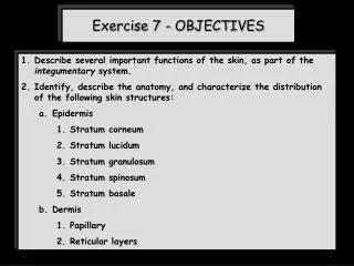
Exercise 7 - OBJECTIVES • Describe several important functions of the skin, as part of the integumentary system. • Identify, describe the anatomy, and characterize the distribution of the following skin structures: • Epidermis • Stratum corneum • Stratum lucidum • Stratum granulosum • Stratum spinosum • Stratum basale • Dermis • 1. Papillary • 2. Reticular layers
Exercise 7 - OBJECTIVES • 2. Identify, describe the anatomy, and characterize the distribution of the following integumentary structures: • a. Hair follicles and hair • b. Sebaceous glands • c. Sweat glands • d. Pacinian corpuscles • Merkel cells and Merkel discs • Meissner’s corpuscles • Compare and contrast the properties of the epidermis to the dermis. • How do sweat glands and sebaceous glands differ? • Compare and contrast the anatomy and function of the eccrine and apocrine sweat glands. • Describe what determines skin color. • Describe the function of melanin.
SKIN LAYERS Epidermis Dermis X Marieb; Fig. 5.1
X http://www.sunyniagara.cc.ny.us/val/histology.html
Major Layers of the Integument White arrows – epidermis Blue arrow - dermis Red arrows – dermal papillary layer (primarily loose connective tissue) Greenarrows – dermal reticular layer (primarily dense irregular connective tissue) Black arrow – hypodermis (superficial fascia) (not part of the “skin”)
SKIN LAYERS Epidermis Dermis Stratum corneum Papillary layer Stratum lucidum (maybe) Reticular layer Stratum granulosum Stratum spinosum Stratum basale
So, name the layers, and define their basic characteristics.
epidermis keratinized stratified squamous epithelium dermis strong, flexible connective tissue
What are the names of the epidermal layers of thin skin? stratum corneum stratum granulosum stratum spinosum stratum basale
Stratum corneum Stratum lucidum Stratum granulosum Stratum spinosum Stratum basale Dermal Papillary Dermal Reticular
Stratum corneum Stratum lucidum Stratum granulosum Stratum spinosum Stratum basale Meissner’s corpuscles
Epidermis of Palm of Hand Greenarrow – epidermis of thick skin Red dashed line – epidermal ridges projecting into the dermis to protect from shear forces on the skin Yellow dashed line – dermal papillae projecting into the epidermis also provided for strength against shear forces
Epidermal Layers of Palm of Hand Black arrows – stratum corneum, where cells have lost their nuclei Green dotten lines – stratum granulosum Yellow arrows – stratum spinosum White arrows– stratum basale (also called stratum germinativum) single row of cells are constantly undergoing mitosis and giving rise to the overlying epidermis
Closer View of Epidermal Layers of Palm of Hand Green arrow – stratum corneum Red arrows – lightly staining stratum lucidum (ONLY PRESENT IN THICK SKIN) White arrows– darkly staining stratum granulosum Yellow arrow – stratum spinosum
Stratum spinosum Stratum basale Meissner’s corpuscle
Close-Up of Stratum Spinosum Red arrows – desmosome “spines”
Primary Epidermal Cells Keratinocytes Melanocytes Langerhans’ Merkel predominant cell type produce keratin connected to one another by desmosomes arise in stratum basale and are “pushed” upward spider-shaped produce melanin that is then trans-ferred to keratinocytes found in deepest layers star-shaped (epidermal dendritic cells) arise from bone marrow and migrate to epidermis macrophages that help immune system shaped like spiky hemisphere functions in sensation located at epidermal-dermal junction
So, name the epidermal cells and define their basic characteristics. keratinocytes Remember, these are the most predominant cell type in the epidermis. They are first produced at the level of the stratum basale, but then get ‘pushed’ to the surface. As they move ‘upwards’, they lose their organelles, and become somewhat hardened. So, the stratum corneum consists of dead keratinocytes that are ready to be sloughed off.
Note how the keratinocytes at this level have lost their nuclei. Pushed to surface In other words, these keratinocytes are anucleated. You should remember this concept from the information provided on the histology of epithelial tissue in Exercise 6A. The next slide is from this Exercise. If this is not familiar information, go back and review all of these slides.
STRATIFIED SQUAMOUS EPITHELIUM OF THE SKIN Greenline – nucleated non-keratinized cells Yellow line – nonnucleated keratinized cells Blue line – depth of entire epithelium
Pigmented keratinocytes http://www.usask.ca/anatomy/teaching/anat232/Integument.jpg/II-55%20Pig.%20keratinocytes%20HP.jpg
So, name the epidermal cells and define their basic characteristics. melanocyte Remember, these are the cells that produce melanin, which is the pigment that protects many of the cells in the epidermis. Once produced, melanin is actually phagocytosed by the keratinocytes. Do you see the “dots” in the keratinocytes that are located near the melanocytes?
melanin-containing keratinocyte being ‘pushed toward the stratum corneum keratinocyte undergoing mitosis AND accumulating melanin
melanocytes http://cal.vet.upenn.edu/histo/skin/melanocytes.html
Melanocyte http://www.usask.ca/anatomy/teaching/anat232/Integument.jpg/II-53%20Melanocyte%20HP.jpg
Melanocyte http://www.usask.ca/anatomy/teaching/anat232/Integument.jpg/II-53%20Melanocyte%20LP.jpg
Melanocyte http://www.usask.ca/anatomy/teaching/anat232/Integument.jpg/II-54%20Melanocyte.jpg
http://www.wtmcgee.com/img/suntan.jpg So you should now be able to explain why a suntan ‘fades’. The keratinocytes that had the melanin are ‘pushed’ to the surface of your skin, and are sloughed off.
Which of these two individuals has a greater number of melanocytes? People of all skin colors have about the same number of melanocytes. Differences in skin color result from differences in the synthesis of melanin and how ‘clumped’ the melanin is within the keratinocytes. http://photos.imageevent.com/dreamkast/rockets/dreaming.jpg
So, name the epidermal cells and define their basic characteristics. Langerhans’ Remember, these are the epidermal dendritic cells. They are phagocytic and play a role in immunity. This should make sense to you. Since the skin is the first structure that is often encountered by a foreign pathogen, we should have a way to defend ourselves at this level.