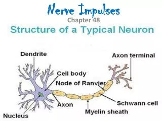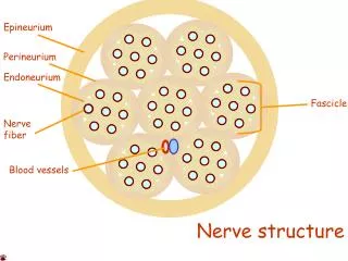
Nerve Fiber Classification
Nerve Fiber Classification. Nerve fibers are classified according to: Diameter Degree of myelination Speed of conduction. Neurons (Nerve Cells). Figure 11.4b. Comparison of Structural Classes of Neurons. Table 11.1.1. Comparison of Structural Classes of Neurons. Table 11.1.2.

Nerve Fiber Classification
E N D
Presentation Transcript
Nerve Fiber Classification • Nerve fibers are classified according to: • Diameter • Degree of myelination • Speed of conduction
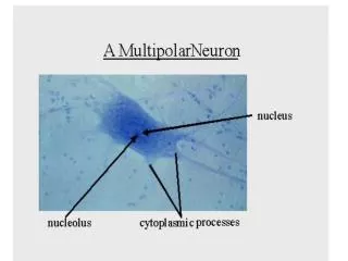
Neurons (Nerve Cells) Figure 11.4b
Comparison of Structural Classes of Neurons Table 11.1.1
Comparison of Structural Classes of Neurons Table 11.1.2
Comparison of Structural Classes of Neurons Table 11.1.3
Changes in Membrane Potential Figure 11.9
Graded Potentials • Short-lived, local changes in membrane potential • Decrease in intensity with distance • Their magnitude varies directly with the strength of the stimulus • Sufficiently strong graded potentials can initiate action potentials • Current is quickly dissipated due to the leaky plasma membrane • Can only travel over short distances
Action Potential Figure 11.15
Absolute Refractory Period Figure 11.15
Absolute Refractory Period • Time from the opening of the Na+ activation gates until the closing of inactivation gates • The absolute refractory period: • Prevents the neuron from generating an action potential • Ensures that each action potential is separate • Enforces one-way transmission of nerve impulses
Relative Refractory Period • The interval following the absolute refractory period when: • Sodium gates are closed • Potassium gates are open • Repolarization is occurring
Myelin Sheath and Neurilemma: Formation Figure 11.5a-c
Saltatory Conduction Figure 11.16
Saltatory Conduction • Current passes through a myelinated axon only at the nodes of Ranvier • Voltage-gated Na+ channels are concentrated at these nodes • Action potentials are triggered only at the nodes and jump from one node to the next • Much faster than conduction along unmyelinated axons
Conduction Velocities of Axons • Conduction velocities vary widely among neurons • Rate of impulse propagation is determined by: • Axon diameter – the larger the diameter, the faster the impulse • Presence of a myelin sheath – myelination dramatically increases impulse speed
Coding for Stimulus Intensity • Upward arrows – stimulus applied • Downward arrows – stimulus stopped • Length of arrows – strength of stimulus Figure 11.14
Coding for Stimulus Intensity • All action potentials are alike and are independent of stimulus intensity • Strong stimuli can generate an action potential more often than weaker stimuli • The CNS determines stimulus intensity by the frequency of impulse transmission
Multiple Sclerosis (MS) • An autoimmune disease that mainly affects young adults • Symptoms include visual disturbances, weakness, loss of muscular control, and urinary incontinence • Nerve fibers are severed and myelin sheaths in the CNS become nonfunctional scleroses • Shunting and short-circuiting of nerve impulses occurs
Multiple Sclerosis: Treatment • The advent of disease-modifying drugs including interferon beta-1a and -1b, Avonex, Betaseran, and Copazone: • Hold symptoms at bay • Reduce complications • Reduce disability