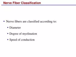
Nerve Fiber Classification
Nerve Fiber Classification. Nerve fibers are classified according to: Diameter Degree of myelination Speed of conduction. Synapses. A junction that mediates information transfer from one neuron: To another neuron To an effector cell

Nerve Fiber Classification
E N D
Presentation Transcript
Nerve Fiber Classification • Nerve fibers are classified according to: • Diameter • Degree of myelination • Speed of conduction
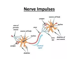
Synapses • A junction that mediates information transfer from one neuron: • To another neuron • To an effector cell • Presynaptic neuron – conducts impulses toward the synapse • Postsynaptic neuron – transmits impulses away from the synapse
Synapses Figure 11.17
Types of Synapses • Axodendritic – synapses between the axon of one neuron and the dendrite of another • Axosomatic – synapses between the axon of one neuron and the soma of another • Other types of synapses include: • Axoaxonic (axon to axon) • Dendrodendritic (dendrite to dendrite) • Dendrosomatic (dendrites to soma)
Electrical Synapses • Electrical synapses: • Are less common than chemical synapses • Correspond to gap junctions found in other cell types • Are important in the CNS in: • Arousal from sleep • Mental attention • Emotions and memory • Ion and water homeostasis
Chemical Synapses • Specialized for the release and reception of neurotransmitters • Typically composed of two parts: • Axonal terminal of the presynaptic neuron, which contains synaptic vesicles • Receptor region on the dendrite(s) or soma of the postsynaptic neuron
Synaptic Cleft • Fluid-filled space separating the presynaptic and postsynaptic neurons • Prevents nerve impulses from directly passing from one neuron to the next • Transmission across the synaptic cleft: • Is a chemical event (as opposed to an electrical one) • Ensures unidirectional communication between neurons
Synaptic Cleft: Information Transfer • Nerve impulses reach the axonal terminal of the presynaptic neuron and open Ca2+ channels • Neurotransmitter is released into the synaptic cleft via exocytosis in response to synaptotagmin • Neurotransmitter crosses the synaptic cleft and binds to receptors on the postsynaptic neuron • Postsynaptic membrane permeability changes, causing an excitatory or inhibitory effect
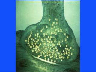
Synaptic Cleft: Information Transfer Neurotransmitter Na+ Ca2+ Axon terminal of presynaptic neuron Action potential Receptor 1 Postsynaptic membrane Mitochondrion Postsynaptic membrane Axon of presynaptic neuron Ion channel open Synaptic vesicles containing neurotransmitter molecules 5 Degraded neurotransmitter 2 Synaptic cleft 3 4 Ion channel closed Ion channel (closed) Ion channel (open) Figure 11.18
Termination of Neurotransmitter Effects • Neurotransmitter bound to a postsynaptic neuron: • Produces a continuous postsynaptic effect • Blocks reception of additional “messages” • Must be removed from its receptor • Removal of neurotransmitters occurs when they: • Are degraded by enzymes • Are reabsorbed by astrocytes or the presynaptic terminals • Diffuse from the synaptic cleft
Synaptic Delay • Neurotransmitter must be released, diffuse across the synapse, and bind to receptors • Synaptic delay – time needed to do this (0.3-5.0 ms) • Synaptic delay is the rate-limiting step of neural transmission
Postsynaptic Potentials • Neurotransmitter receptors mediate changes in membrane potential according to: • The amount of neurotransmitter released • The amount of time the neurotransmitter is bound to receptors • The two types of postsynaptic potentials are: • EPSP – excitatory postsynaptic potentials • IPSP – inhibitory postsynaptic potentials
Excitatory Postsynaptic Potentials • EPSPs are graded potentials that can initiate an action potential in an axon • Use only chemically gated channels • Na+ and K+ flow in opposite directions at the same time • Postsynaptic membranes do not generate action potentials
Excitatory Postsynaptic Potential (EPSP) Figure 11.19a
Inhibitory Synapses and IPSPs • Neurotransmitter binding to a receptor at inhibitory synapses: • Causes the membrane to become more permeable to potassium and chloride ions • Leaves the charge on the inner surface negative • Reduces the postsynaptic neuron’s ability to produce an action potential
Inhibitory Postsynaptic (IPSP) Figure 11.19b
Summation • A single EPSP cannot induce an action potential • EPSPs must summate temporally or spatially to induce an action potential • Temporal summation – presynaptic neurons transmit impulses in rapid-fire order
Summation • Spatial summation – postsynaptic neuron is stimulated by a large number of terminals at the same time • IPSPs can also summate with EPSPs, canceling each other out
Summation Figure 11.20
Neurotransmitters • Chemicals used for neuronal communication with the body and the brain • 50 different neurotransmitters have been identified • Classified chemically and functionally
Chemical Neurotransmitters • Acetylcholine (ACh) • Biogenic amines • Amino acids • Peptides • Novel messengers: ATP and dissolved gases NO and CO
Neurotransmitters: Acetylcholine • First neurotransmitter identified, and best understood • Released at the neuromuscular junction • Synthesized and enclosed in synaptic vesicles
Neurotransmitters: Acetylcholine • Degraded by the enzyme acetylcholinesterase (AChE) • Released by: • All neurons that stimulate skeletal muscle • Some neurons in the autonomic nervous system
Neurotransmitters: Biogenic Amines • Include: • Catecholamines – dopamine, norepinephrine (NE), and epinephrine • Indolamines – serotonin and histamine • Broadly distributed in the brain • Play roles in emotional behaviors and our biological clock
Synthesis of Catecholamines • Enzymes present in the cell determine length of biosynthetic pathway • Norepinephrine and dopamine are synthesized in axonal terminals • Epinephrine is released by the adrenal medulla Figure 11.21
Neurotransmitters: Amino Acids • Include: • GABA – Gamma ()-aminobutyric acid • Glycine • Aspartate • Glutamate • Found only in the CNS
Neurotransmitters: Peptides • Include: • Substance P – mediator of pain signals • Beta endorphin, dynorphin, and enkephalins • Act as natural opiates; reduce pain perception • Bind to the same receptors as opiates and morphine • Gut-brain peptides – somatostatin, and cholecystokinin
Neurotransmitters: Novel Messengers • ATP • Is found in both the CNS and PNS • Produces excitatory or inhibitory responses depending on receptor type • Induces Ca2+ wave propagation in astrocytes • Provokes pain sensation
Neurotransmitters: Novel Messengers • Nitric oxide (NO) • Activates the intracellular receptor guanylyl cyclase • Is involved in learning and memory • Carbon monoxide (CO) is a main regulator of cGMP in the brain
Functional Classification of Neurotransmitters • Two classifications: excitatory and inhibitory • Excitatory neurotransmitters cause depolarizations (e.g., glutamate) • Inhibitory neurotransmitters cause hyperpolarizations (e.g., GABA and glycine)
Functional Classification of Neurotransmitters • Some neurotransmitters have both excitatory and inhibitory effects • Determined by the receptor type of the postsynaptic neuron • Example: acetylcholine • Excitatory at neuromuscular junctions with skeletal muscle • Inhibitory in cardiac muscle
Neurotransmitter Receptor Mechanisms • Direct: neurotransmitters that open ion channels • Promote rapid responses • Examples: ACh and amino acids • Indirect: neurotransmitters that act through second messengers • Promote long-lasting effects • Examples: biogenic amines, peptides, and dissolved gases
Channel-Linked Receptors • Composed of integral membrane protein • Mediate direct neurotransmitter action • Action is immediate, brief, simple, and highly localized • Ligand binds the receptor, and ions enter the cells • Excitatory receptors depolarize membranes • Inhibitory receptors hyperpolarize membranes
Channel-Linked Receptors Figure 11.22a
G Protein-Linked Receptors • Responses are indirect, slow, complex, prolonged, and often diffuse • These receptors are transmembrane protein complexes • Examples: muscarinic ACh receptors, neuropeptides, and those that bind biogenic amines
G Protein-Linked Receptors: Mechanism • Neurotransmitter binds to G protein-linked receptor • G protein is activated and GTP is hydrolyzed to GDP • The activated G protein complex activates adenylate cyclase
G Protein-Linked Receptors: Mechanism • Adenylate cyclase catalyzes the formation of cAMP from ATP • cAMP, a second messenger, brings about various cellular responses
Neurotransmitter Receptor Mechanism Ions flow Blocked ion flow Ion channel Adenylate cyclase Channel closed Channel open (a) Neurotransmitter (ligand) released from axon terminal of presynaptic neuron PPi 4 GTP Changes in membrane permeability and potential 5 3 cAMP 1 ATP 5 3 GTP Protein synthesis Enzyme activation 2 GDP GTP Receptor Activation of specific genes G protein (b) Nucleus Figure 11.22b
G Protein-Linked Receptors: Effects • G protein-linked receptors activate intracellular second messengers including Ca2+, cGMP, diacylglycerol, as well as cAMP • Second messengers: • Open or close ion channels • Activate kinase enzymes • Phosphorylate channel proteins • Activate genes and induce protein synthesis
Neural Integration: Neuronal Pools • Functional groups of neurons that: • Integrate incoming information • Forward the processed information to its appropriate destination
Neural Integration: Neuronal Pools • Simple neuronal pool • Input fiber – presynaptic fiber • Discharge zone – neurons most closely associated with the incoming fiber • Facilitated zone – neurons farther away from incoming fiber
Simple Neuronal Pool Figure 11.23
Types of Circuits in Neuronal Pools • Divergent – one incoming fiber stimulates ever increasing number of fibers, often amplifying circuits Figure 11.24a, b
Types of Circuits in Neuronal Pools • Convergent – opposite of divergent circuits, resulting in either strong stimulation or inhibition Figure 11.24c, d
Types of Circuits in Neuronal Pools • Reverberating – chain of neurons containing collateral synapses with previous neurons in the chain Figure 11.24e
Types of Circuits in Neuronal Pools • Parallel after-discharge – incoming neurons stimulate several neurons in parallel arrays Figure 11.24f
Patterns of Neural Processing • Serial Processing • Input travels along one pathway to a specific destination • Works in an all-or-none manner • Example: spinal reflexes
Patterns of Neural Processing • Parallel Processing • Input travels along several pathways • Pathways are integrated in different CNS systems • One stimulus promotes numerous responses • Example: a smell may remind one of the odor and associated experiences
Development of Neurons • The nervous system originates from the neural tube and neural crest • The neural tube becomes the CNS • There is a three-phase process of differentiation: • Proliferation of cells needed for development • Migration – cells become amitotic and move externally • Differentiation into neuroblasts
Axonal Growth • Guided by: • Scaffold laid down by older neurons • Orienting glial fibers • Release of nerve growth factor by astrocytes • Neurotropins released by other neurons • Repulsion guiding molecules • Attractants released by target cells