Download
1 / 38
400 likes | 696 Vues
Protection, Support, and Movement. Chapter 37. Husky Adaptations. Huskies are adapted to load pulling and long distance running in cold climates Their integument, musculature, and skeleton are specialized for this way of life. Integumentary System. An animal’s outer covering Examples

E N D
Protection, Support, and Movement Chapter 37
Husky Adaptations • Huskies are adapted to load pulling and long distance running in cold climates • Their integument, musculature, and skeleton are specialized for this way of life
Integumentary System • An animal’s outer covering • Examples • Chitin-hardened cuticle of many invertebrates • Vertebrate skin and its derivatives
Vertebrate Skin • Two layers • Upper epidermis • Lower dermis • Lies atop a layer of hypodermis Figure 37.3Page 646
Functions of Human Skin • Protects the body from injury, dehydration, UV radiation, and some pathogens • Helps control temperature • Receives some external stimuli • Produces vitamin D
Sunlight Damages Skin • UV light stimulates melanin production in skin; produces a tan • Tan is the body’s way of protecting itself against UV • Prolonged sun exposure causes elastin fibers to clump, skin to age prematurely
Langerhans Cells • White blood cells that arise in bone marrow, migrate to epidermis • Engulf pathogens and alert immune system • UV radiation can damage these cells and weaken body’s first line of defense
Granstein Cells • Also occur in epidermis • Interact with cells that carry out immune response • Issue suppressor signals that keep immune response under control • Less vulnerable to UV damage than Langerhans cells
Hydrostatic Skeleton • Muscles work against an internal body fluid and redistribute it within a confined space Radial cells are relaxed; longitudinal ones contracted Radial cells are contracted; longitudinal ones relaxed
wing Exoskeleton pivot point Dorsal-ventral muscles contract, exoskeleton pops out, wings move up • Rigid, external body parts receive the applied force of muscle contraction Dorsal-ventral muscles relax, exoskeleton pops back, wings move down In-text figurePage 649
Endoskeleton • All vertebrates • Fins or limbs attach to skeleton at pectoral and pelvic girdles Generalized mammal Figure 37.10cPage 650 pelvic girdle pectoral girdle
Functions of Bone • Interact with muscle to enable movement • Support and anchor muscles • Enclose and protect internal organs • Store calcium and phosphorus • Produce blood cells
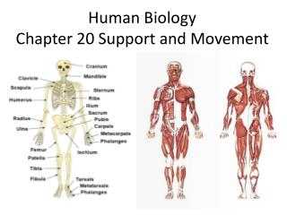
SKULL PECTORAL GIRDLES AND UPPER EXTREMITIES cranial bones facial bones clavicle RIB CAGE scapula sternum humerus ribs radius ulna VERTEBRAL COLUMN vertebrae phalanges carpals intervertebral disks metacarpals PELVIC GIRDLE AND LOWER EXTREMITIES pelvic girdle femur patella tibia fibula tarsals phalanges metatarsals Human Skeleton Stepped Art Figure 37.11Page 651
Long Bone Structure • Compact bone • Spongy bone • Central cavity contains yellow marrow nutrient canal contains yellow marrow compact bone tissue spongy bone tissue Fig. 37.12 (1)Page 652
Compact Bone Structure • Mature compact bone consists of many cylindrical Haversian systems Haversian system blood vessel outer layer of dense connective tissue spongy bonetissue Fig. 37.12 (2)Page 652 compact bone tissue
Bone Marrow • Yellow marrow • Fills the cavities of adult long bones • Is largely fat • Red marrow • Occurs in spongy bone of some bones • Produces blood cells
Bone Remodeling • In adults, bone building and bone breakdown continue constantly • Osteoblasts deposit bone • Osteoclasts secrete enzymes that degrade it • Remodeling adjusts bone strength and helps maintain blood calcium levels
Bone Density • Exercise can increase bone density • Osteoporosis is a decrease in bone density • May occur when the action of osteoclasts outpaces that of osteoblasts • May also occur as a result of inability to absorb calcium
Joints • Areas of contact or near contact between bones • Fibrous joints • Short connecting fibers join bones • Synovial joints • Move freely; ligaments connect bones • Cartilaginous joints • Straps of cartilage allow slight movement
Tendons Attach Muscle to Bone muscle tendon bursae synovial cavity Figure 37.15Page 654
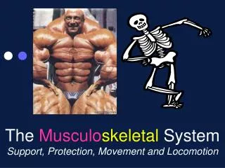
Skeletal Muscle • Bundles of striped muscle cells • Attaches to bone • Often works in opposition biceps triceps Figure 37.17Page 654
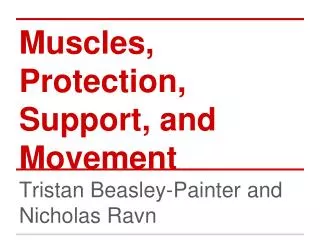
triceps brachii biceps brachii Major Human Muscles deltoid pectoralis major trapezius serratus anterior external oblique latissimus dorsi rectus abdominus gluteus maximus adductor longus biceps femoris sartorius quadriceps femoris gastrocnemius tibialis anterior Figure 37.18Page 655
Skeletal Muscle Structure • A muscle is made up of muscle cells • A muscle fiber is a single muscle cell • Each fiber contains many myofibrils myofibril Figure 37.19aPage 656
Sarcomere A myofibril is made up of thick and thin filaments arranged in sarcomeres sarcomere sarcomere sarcomere sarcomere Z line Z line Z line Figure 37.19bPage 656
Muscle Microfilaments Thin filaments • Like two strands of pearls twisted together • Pearls are actin • Other proteins in grooves in filament Thick filaments • Composed of myosin • Each myosin molecule has tail and a double head Figure 37.19cPage 656
Sliding-Filament Model • Myosin heads attach to actin filaments • Myosin heads tilt toward sarcomere center, pulling actin with them Fig. 37.20c-gPage 657
Sliding-Filament Model Sarcomere shortens because the actin filaments are pulled inward, toward the sarcomere center Fig. 37.20a,bPage 657
Nervous System Controls Contraction • Signals from nervous system travel along spinal cord, down a motor neuron • Endings of motor neuron synapse on a muscle cell at a neuromuscular junction
Role of Calcium in Contraction • T tubules in the sarcoplasmic reticulum relay signal • Calcium ions are released
Troponin and Tropomyosin • Lie in groove in actin filament • When muscle is relaxed, tropomyosin blocks myosin binding site troponin myosin binding site blocked actin Figure 37.22Page 659
Troponin and Tropomyosin • When troponin binds calcium ions, it changes shape and moves tropomyosin • Cross-bridge formation and contraction can now proceed myosinhead actin Figure 37.22Page 659
Contraction Requires Energy • Muscle cells require huge amounts of ATP energy to power contraction • The cells have only a very small store of ATP • Three pathways supply ATP to power muscle contraction
ATP for Contraction ADP + Pi Pathway 1 Dephosphorylation Creatine Phosphate relaxation contraction creatine Pathway 2 Aerobic Respiration Pathway 3 Glycolysis Alone glucose from bloodstream and from glycogen break down in cells oxygen Figure 37.23Page 659
Muscle Tension • Mechanical force a contracting muscle exerts on an object • For a muscle to shorten, muscle tension must exceed the load that opposes it • The load may be the weight of an object or gravity’s pull on the muscle
Two Types of Contraction contracted muscle can’t shorten contracted muscle can shorten isotonic contraction isometric contraction Figure 37.24Page 660
Motor Unit • One neuron and all the muscle cells that form junctions with its endings • When a motor neuron is stimulated, all the muscle cells it supplies are activated to contract simultaneously • Each muscle consists of many motor units
peak relaxation Twitches and Tetanus stimulus contraction starts time number of stimuli per second number of stimuli per second tetanic contraction Figure 37.25Page 660 twitch repeated stimulation
Muscle Fatigue • An inability to maintain muscle tension • Occurs after a period of tetanic contraction • Different types of muscle show different fatigue patterns