Download
1 / 43
430 likes | 570 Vues
Concept 49.5: Animal skeletons function in support, protection, and movement The various types of animal movements All result from muscles working against some type of skeleton. Types of Skeletons. The three main functions of a skeleton are Support, protection, and movement

E N D
Concept 49.5: Animal skeletons function in support, protection, and movement • The various types of animal movements • All result from muscles working against some type of skeleton
Types of Skeletons • The three main functions of a skeleton are • Support, protection, and movement • The three main types of skeletons are • Hydrostatic skeletons, exoskeletons, and endoskeletons
Hydrostatic Skeletons • A hydrostatic skeleton • Consists of fluid held under pressure in a closed body compartment • This is the main type of skeleton • In most cnidarians, flatworms, nematodes, and annelids
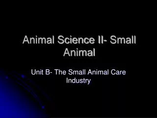
Circular muscle contracted Longitudinal muscle relaxed (extended) Circular muscle relaxed Longitudinal muscle contracted (a) Body segments at the head and just in front of the rear are short and thick (longitudinal muscles contracted; circular muscles relaxed) and anchored to the ground by bristles. The other segments are thin and elongated (circular muscles contracted; longitudinal muscles relaxed.) Head Bristles (b) The head has moved forward because circular muscles in the head segments have contracted. Segments behind the head and at the rear are now thick and anchored, thus preventing the worm from slipping backward. (c) The head segments are thick again and anchored in their new positions. The rear segments have released their hold on the ground and have been pulled forward. • Annelids use their hydrostatic skeleton for peristalsis • A type of movement on land produced by rhythmic waves of muscle contractions Figure 49.25a–c
Exoskeletons • An exoskeleton is a hard encasement • Deposited on the surface of an animal • Exoskeletons • Are found in most molluscs and arthropods
Endoskeletons • An endoskeleton consists of hard supporting elements • Such as bones, buried within the soft tissue of an animal • Endoskeletons • Are found in sponges, echinoderms, and chordates
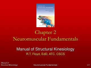
The mammalian skeleton is built from more than 200 bones • Some fused together and others connected at joints by ligaments that allow freedom of movement
Head ofhumerus key Examplesof joints Axial skeleton Skull Appendicularskeleton Scapula 1 Clavicle Shouldergirdle Scapula Sternum 1Ball-and-socket joints, where the humerus contactsthe shoulder girdle and where the femur contacts thepelvic girdle, enable us to rotate our arms andlegs and move them in several planes. Rib 2 Humerus 3 Vertebra Radius Ulna Humerus Pelvicgirdle Carpals Ulna Phalanges 2Hinge joints, such as between the humerus andthe head of the ulna, restrict movement to a singleplane. Metacarpals Femur Patella Tibia Fibula Ulna Radius Tarsals 3Pivot joints allow us to rotate our forearm at theelbow and to move our head from side to side. Metatarsals Phalanges • The human skeleton Figure 49.26
Physical Support on Land • In addition to the skeleton • Muscles and tendons help support large land vertebrates
Concept 49.6: Muscles move skeletal parts by contracting • The action of a muscle • Is always to contract
Human Grasshopper Extensormusclerelaxes Bicepscontracts Tibiaflexes Flexormusclecontracts Tricepsrelaxes Forearmflexes Extensormusclecontracts Tibiaextends Bicepsrelaxes Forearmextends Flexormusclerelaxes Triceps contracts • Skeletal muscles are attached to the skeleton in antagonistic pairs • With each member of the pair working against each other Figure 49.27
Muscle Bundle ofmuscle fibers Nuclei Single muscle fiber (cell) Plasma membrane Myofibril Z line Lightband Dark band Sarcomere TEM 0.5 m A band I band I band M line Thickfilaments(myosin) Thinfilaments(actin) H zone Z line Z line Sarcomere Vertebrate Skeletal Muscle • Vertebrate skeletal muscle • Is characterized by a hierarchy of smaller and smaller units animation Figure 49.28
A skeletal muscle consists of a bundle of long fibers • Running parallel to the length of the muscle • A muscle fiber (muscle cell) • Is itself a bundle of smaller myofibrils arranged longitudinally
The myofibrils are composed to two kinds of myofilaments • Thin filaments, consisting of two strands of actin and one strand of regulatory protein • Thick filaments, staggered arrays of myosin molecules
Skeletal muscle is also called striated muscle • Because the regular arrangement of the myofilaments creates a pattern of light and dark bands
Each repeating unit is a sarcomere • Bordered by Z lines • The areas that contain the myofilments • Are the I band, A band, and H zone
The Sliding-Filament Model of Muscle Contraction • According to the sliding-filament model of muscle contraction • The filaments slide past each other longitudinally, producing more overlap between the thin and thick filaments
0.5 m (a) Relaxed muscle fiber. In a relaxed muscle fiber, the I bandsand H zone are relatively wide. Z H A Sarcomere (b) Contracting muscle fiber. During contraction, the thick andthin filaments slide past each other, reducing the width of theI bands and H zone and shortening the sarcomere. (c) Fully contracted muscle fiber. In a fully contracted musclefiber, the sarcomere is shorter still. The thin filaments overlap,eliminating the H zone. The I bands disappear as the ends ofthe thick filaments contact the Z lines. • As a result of this sliding • The I band and the H zone shrink Figure 49.29a–c
The sliding of filaments is based on • The interaction between the actin and myosin molecules of the thick and thin filaments • The “head” of a myosin molecule binds to an actin filament • Forming a cross-bridge and pulling the thin filament toward the center of the sarcomere
Thick filament Thin filaments 1 Starting here, the myosin head is bound to ATP and is in its low-energy confinguration. 5 Binding of a new mole- cule of ATP releases the myosin head from actin, and a new cycle begins. Thin filament Myosin head (low-energy configuration) The myosin head hydrolyzes ATP to ADP and inorganic phosphate ( I ) and is in its high-energy configuration. ATP 2 ATP Cross-bridge binding site Thick filament P Actin Thin filament moves toward center of sarcomere. Myosin head (high-energy configuration) ADP Myosin head (low-energy configuration) P i 1 The myosin head binds toactin, forming a cross-bridge. 3 ADP + Cross-bridge ADP P i P i Releasing ADP and ( i), myosinrelaxes to its low-energy configuration, sliding the thin filament. 4 P • Myosin-actin interactions underlying muscle fiber contraction Figure 49.30
The Role of Calcium and Regulatory Proteins • A skeletal muscle fiber contracts • Only when stimulated by a motor neuron
Tropomyosin Ca2+-binding sites Actin Troponin complex (a) Myosin-binding sites blocked • When a muscle is at rest • The myosin-binding sites on the thin filament are blocked by the regulatory protein tropomyosin Figure 49.31a
Ca2+ Myosin-binding site (b) Myosin-binding sites exposed • For a muscle fiber to contract • The myosin-binding sites must be uncovered • This occurs when calcium ions (Ca2+) • Bind to another set of regulatory proteins, the troponin complex Figure 49.31b
Motorneuron axon Mitochondrion Synapticterminal T tubule Sarcoplasmicreticulum Ca2+ releasedfrom sarcoplasmicreticulum Myofibril Sarcomere Plasma membraneof muscle fiber • The stimulus leading to the contraction of a skeletal muscle fiber • Is an action potential in a motor neuron that makes a synapse with the muscle fiber Figure 49.32
The synaptic terminal of the motor neuron • Releases the neurotransmitter acetylcholine, depolarizing the muscle and causing it to produce an action potential
Action potentials travel to the interior of the muscle fiber • Along infoldings of the plasma membrane called transverse (T) tubules • The action potential along the T tubules • Causes the sarcoplasmic reticulum to release Ca2+ • The Ca2+ binds to the troponin-tropomyosin complex on the thin filaments • Exposing the myosin-binding sites and allowing the cross-bridge cycle to proceed
Acetylcholine (ACh) released by synaptic terminal diffuses across synapticcleft and binds to receptor proteins on muscle fiber’s plasma membrane, triggering an action potential in muscle fiber. Synapticterminalof motorneuron 1 PLASMA MEMBRANE Synaptic cleft T TUBULE Action potential is propa- gated along plasma membrane and down T tubules. 2 ACh SR 4 Action potential triggers Ca2+ release from sarco- plasmic reticulum (SR). 3 Ca2 Calcium ions bind to troponin; troponin changes shape, removing blocking action of tropomyosin; myosin-binding sites exposed. Tropomyosin blockage of myosin- binding sites is restored; contraction ends, and muscle fiber relaxes. 7 Ca2 CYTOSOL Cytosolic Ca2+ is removed by active transport into SR after action potential ends. 6 ADP P2 Myosin cross-bridges alternately attach to actin and detach, pulling actin filaments toward center of sarcomere; ATP powers sliding of filaments. 5 • Review of contraction in a skeletal muscle fiber Figure 49.33
Neural Control of Muscle Tension • Contraction of a whole muscle is graded • Which means that we can voluntarily alter the extent and strength of its contraction
There are two basic mechanisms by which the nervous system produces graded contractions of whole muscles • By varying the number of fibers that contract • By varying the rate at which muscle fibers are stimulated
Motorunit 1 Motorunit 2 Spinal cord Synaptic terminals Nerve Motor neuroncell body Motor neuronaxon Muscle Muscle fibers Tendon • In a vertebrate skeletal muscle • Each branched muscle fiber is innervated by only one motor neuron • Each motor neuron • May synapse with multiple muscle fibers Figure 49.34
A motor unit • Consists of a single motor neuron and all the muscle fibers it controls • Recruitment of multiple motor neurons • Results in stronger contractions
Tetanus Tension Summation of two twitches Singletwitch Time Actionpotential Pair ofactionpotentials Series of action potentials at high frequency • A twitch • Results from a single action potential in a motor neuron • More rapidly delivered action potentials • Produce a graded contraction by summation Figure 49.35
Tetanus is a state of smooth and sustained contraction • Produced when motor neurons deliver a volley of action potentials
Types of Muscle Fibers • Skeletal muscle fibers are classified as slow oxidative, fast oxidative, and fast glycolytic • Based on their contraction speed and major pathway for producing ATP
Other Types of Muscle • Cardiac muscle, found only in the heart • Consists of striated cells that are electrically connected by intercalated discs • Can generate action potentials without neural input
In smooth muscle, found mainly in the walls of hollow organs • The contractions are relatively slow and may be initiated by the muscles themselves • In addition, contractions may be caused by • Stimulation from neurons in the autonomic nervous system
Concept 49.7: Locomotion requires energy to overcome friction and gravity • Movement is a hallmark of all animals • And usually necessary for finding food or evading predators • Locomotion • Is active travel from place to place
Swimming • Overcoming friction • Is a major problem for swimmers • Overcoming gravity is less of a problem for swimmers • Than for animals that move on land or fly
Locomotion on Land • Walking, running, hopping, or crawling on land • Requires an animal to support itself and move against gravity
Diverse adaptations for traveling on land • Have evolved in various vertebrates Figure 49.36
Flying • Flight requires that wings develop enough lift • To overcome the downward force of gravity
EXPERIMENT Physiologists typically determine an animal’s rate of energy use during locomotion by measuring its oxygen consumption or carbon dioxide production while it swims in a water flume, runs on a treadmill, or flies in a wind tunnel. For example, the trained parakeet shown below is wearing a plastic face mask connected to a tube that collects the air the bird exhales as it flies. RESULTS This graph compares the energy cost, in joules per kilogram of body mass per meter traveled, for animals specialized for running, flying, and swimming (1 J = 0.24 cal). Notice that both axes are plotted on logarithmic scales. Flying Running 102 10 Energy cost (J/Kg/m) 1 Swimming 10–1 10–3 1 103 106 Body mass(g) Comparing Costs of Locomotion • The energy cost of locomotion • Depends on the mode of locomotion and the environment CONCLUSION For animals of a given body mass, swimming is the most energy-efficient and running the least energy-efficient mode of locomotion. In any mode, a small animal expends more energy per kilogram of body mass than a large animal. CONCLUSION Figure 49.37