The Breast: an Overview
670 likes | 843 Vues
The Breast: an Overview. Lisa S. Dresner, MD, FACS Associate Professor of Surgery SUNY Downstate. Prevalence/Incidence. 200,000 new cases in USA / year Incidence 121 / 100,000 white women 99 / 100,000 black women Stage Increased numbers of early and non-invasive cancers

The Breast: an Overview
E N D
Presentation Transcript
The Breast: an Overview Lisa S. Dresner, MD, FACS Associate Professor of Surgery SUNY Downstate
Prevalence/Incidence • 200,000 new cases in USA / year • Incidence • 121 / 100,000 white women • 99 / 100,000 black women • Stage • Increased numbers of early and non-invasive cancers • Stable or slightly decreased number of advanced • Rates: vary geographically and ethnically • Rates vary greatly by age
Risk of Breast Cancer Lifetime risk of dx: 13.22 % Lifetime risk of dying: 2.96 %
Physiology • Cell Regulation: • Growth development and function under hormone control • Binding of hormone to specific cell receptors trigger effects • Estrogens: • important in development, growth and differentiation. Normal and most malignant breast cells contain ER receptors. • E-ER complex binds with nuclear chromatin and influences protein production including progesterone receptor (PR)
History: • Complaint, ask about SBE • Timing and nature of previous breast surgery (atypia, cancer etc) • Family history of breast or ovarian cancer • Use of hormones • Reproductive history • Radiation exposure
Physical Exam • Best/easiest during week after menses • Palpate supraclavicular, cervical and axillary nodes • Skin changes: dimpling, edema, nipple change • With patient supine with hand over head examine breast in a systematic way against the chest wall
Evaluation of Breast Mass • In women under 30 ultrasound • In women over 30 mammo±ultrasound • As a rule all except obviously benign masses should have pathological diagnosis • Open biopsy • Core biopsy • FNA • Ultrasound guided core biopsy (highly sensitive and specific) • If the mass is indeterminate by your exam consider ultrasound to confirm • If mass not palpable stereotactic core biopsy
Screening: • No controversy: all women aged 50 and older should have a mammogram every 1-2 years as well as an annual clinical breast exam (CBE) • Women 40-50: guidelines ACS mammogram every 1-2 years as well as an annual clinical breast exam (CBE) • High Risk: earlier mammography.
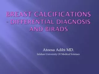
Mammogram: ACR Classification Standardized way of reporting mammogram results.
Masses: Round Circumscribed Microlobulated Oval Obscured Lobulated Ill-defined Irregular Spiculated
Management of Non-Palpable Mammographic abnormalities • Ultrasound: is there a mass? • Ultrasound guided core biopsy may be diagnostic • Stereotactic core biopsy • Mammographic abnormalities • Mammotome (mammo-guided very big core; may be excisional) • Needle localization biopsy • Mammo or ultrasound guided open biopsy • Cryoablation: for bx proven benign
MRI for evaluation of the breast • Highly sensative but high false postive rate • Useful for screening BRCA patients • May be useful in staging known breast cancer • May become an important screening modality
Other imaging modalities • Tc99m sestamibi scan (Miraluma) • Tomosynthesis (variation of mammogram)
MRI • Extremely sensitive (?high false positives?) • May be useful in staging • May be useful in high risk patients with difficult mammograms • Not yet approved for screening
Benign Breast Disorders: 1 • Fibrocystic “disease” • Nodular, lumpy, tender breasts: • Mastodynia • Clear/milky nipple discharge • Within the range of normal • Confirm benign-ness, Reassurance, symptomatic relief. Encourage BSE • Fibrocystic features • Adenosis, cysts, fibrosis (not increased risk) • Ductal and lobular hyperplasia with or without atypia (with increased risk)
Breast cysts: • A palpable mass could be a cyst • Simple cysts need no treatment • Needle aspiration to confirm, or for pain relief • Ultrasound (conclusive) • Complex cysts, bloody cysts deserve evaluation and biopsy (open or ultrasound guided core) • Excision if diagnosis is in doubt after minimal invasive biopsy
Fibroadenoma • May present at any age but most common women 16-24. • Rubbery, mobile, well defined • Confirm by core, excision, FNA, or ultrasound, and/or short interval observation by ultrasound • Giant fibroadenomas: may be very large and grow rapidly (late teens and perimenopause): RX: enucleation • Actual pathology may be adenoma, fibroadenoma,etc
Phylloides Tumor • Old name cystosarcoma phylloides • Mesenchymal tumor: leaf like masses, cellular with necrosis and hemorrhage • May occur in adolescent (generally benign) or premenopausal woman (may be malignant) • Treated with excision with margins • 25% risk of local recurrence in 10 years even with ‘benign” path • Mitotic figure count is one predictor of malignancy • Metastasis even in “malignant” tumors are rare • Younger: more likely benign, older women more likely malignant
Other benign breast masses • Sclerosing adenosis • Radial scar • Fat necrosis • Ductal ectasia • Lactational mastitis and galactocele • Mondor’s disease • Intraductal papilloma • Lactating adenoma
Mastodynia • Cyclical or continuous. May be referred to axilla, upper arm, may improve with menopause • Rarely associated with malignancy • Continuous: may be related to a large cyst,infection or inflammation • Reassurance, NSAIDS, well fitted brassiere, caffeine reduction, evening primrose oil, cessation of tobacco use (takes months) • Danazol, bromocriptine and tamoxifen (side effects prohibitive) • ?SSRI
Nipple Discharge • Most common after lactation (as long as 2 years) • Subareolar infection (increased risk in smokers) • Galactorrhea (bilateral, milky) prolactin excess • Fibrocystic: green, yellow, brown (guiac) • Bloody: intraductal papilloma (benign), Cancer should be ruled out. Ductogram (galactogram) may be helpful
Hyperplasias: not malignant but not really benign either • Ductal hyperplasias • Mild • Moderate • Florid • Atypical Ductal hyperplasia (ADH) • (Ductal carcinoma in-situ- DCIS*) • Lobular hyperplasias • Lobular hyperplasia • Lobular carcinoma in-situ
Lobular Carcinoma In-situ LCIS Bystander lesion- marker of risk • Commonly occurs in 4th decade of life, 2/3 are premenopausal • Lobular tumors are more likely ER/PR positive • Diagnosis incidental on biopsy of other pathology • Significant life time risk of breast cancer (5.9 to 12 times higher) but the risk is in both breasts • Risk is greater 15-20 years after diagnosis than the immediate post diagnostic period
Lobular Carcinoma • Clinical features, epidemiology and risk factors and treatment not different • Doesn’t form microcalcifications and is extensively infiltrative so may be mammographically occult • May present as “architectural distortion on mamography
Invasive Ductal Carcinoma • Most common tumor: from ductal elements • Invasion of nerves, vessels, lymphatics in the breast parenchyma at edge of lesions may be present and carries a poorer prognosis • May have all or partial characteristics of other types (colloid, tubular, medullary)
Breast Cancer Risk Factors • Greatly increased risk RR>4.0 • Inherited genetic mutations for breast cancer • ≥ 2 first degree relatives with breast cancer diagnosed at early age • Personal history of breast cancer • Age >65 (increasing risk with increasing age to 80)
Breast Cancer Risk Factors • Moderately increased risk factors RR 2.1-4.0 • One first degree relative with breast cancer • Nodular densities on mammogram (>75% of volume) • Atypical hyperplasia on breast biopsy • High dose ionizing radiation to chest
Breast Cancer Risk Factors 3 Low increased risk: RR 1.1-2 High socioeconomic status, urban residence, Northern USA Early menarche (<12), late menopause (>55) No full term pregnancy, late (>30) first term pregnancy Never breast fed Postmenopausal obesity Etoh,consumption HRT, recent oca use Tall Personal history of ca endometrium, ovary or colon Jewish heritage, mammographically dense breasts
Inherited Breast Cancer Syndromes • 1. Li-Fraumeni syndrome: p53 mutation • 2. Mutation on the sht arm of chromosome 2 • 3. BRCA-1 long arm chromosome 17 (associated with breast and ovarian cancer) • 4. BRCA-2 small region of 13q12-13 • Recommendations vary from bilateral salpingo-oophorectomy and prophylactic mastectomy to increased surveillance • Value of SERM (tamoxifen) unclear as most hereditary-linked breast cancers are ER/PR negative
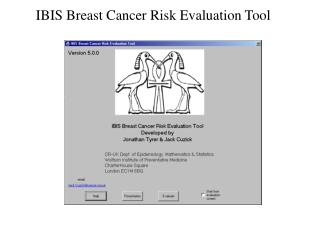
Estimating Risk • Gail Model • calculates risk using 6 key risk factors • Age • Age menarche • Age first birth • Family history (1° female relative) • Number of previous breast biopsies • Number of biopsies with atypical hyperplasia • http://bcra.nci.nih.gov/brc/
Inflammatory breast cancer • Diagnosis: clinical findings of inflamed breast with underlying malignancy. • 35% have obvious mets at time of diagnosis • Mammogram: edema • Dermal or core biopsy • Treatment is neoadjuvant chemotherapy first then mastectomy plus RT
Staging • Primary tumor • Tis: Carcinoma in-situ • T1 : 2 cm or less • T2 : >2 but not more than 5 cm • T3 : >5 cm • T4 : any size with chest wall extension, skin involvement, skin nodules, or inflammatory breast cancer