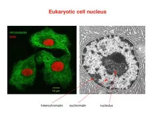

Cellular Structures and Sizes in Microscopy
E N D
Presentation Transcript
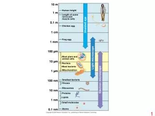
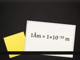
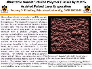
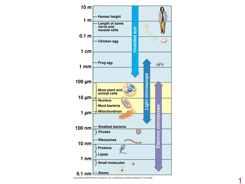
10 m Human height 1 m Length of some nerve and muscle cells 0.1 m Unaided eye Chicken egg 1 cm Frog egg 1 mm 100 µm Most plant and animal cells Light microscope 10 µm Nucleus Most bacteria Mitochondrion 1 µm Electron microscope Smallest bacteria 100 nm Viruses Ribosomes 10 nm Proteins Lipids 1 nm Small molecules Atoms 0.1 nm
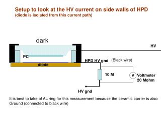
Fimbriae Nucleoid Ribosomes Plasma membrane Cell wall Bacterial chromosome Capsule 0.5 µm Flagella (a) A typical rod-shaped bacterium (b) A thin section through the bacterium Bacillus coagulans (TEM)
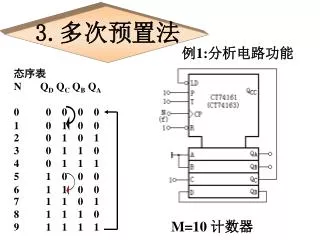
(a) TEM of a plasma membrane Outside of cell Inside of cell 0.1 µm Carbohydrate side chain Hydrophilic region Hydrophobic region Hydrophilic region Phospholipid Proteins (b) Structure of the plasma membrane
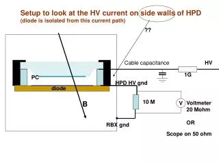
Surface area increases while total volume remains constant 5 1 1 Total surface area [Sum of the surface areas (height width) of all boxes sides number of boxes] 150 750 6 Total volume [height width length number of boxes] 1 125 125 Surface-to-volume (S-to-V) ratio [surface area ÷ volume] 6 1.2 6
Nuclear envelope ENDOPLASMIC RETICULUM (ER) NUCLEUS Nucleolus Rough ER Smooth ER Flagellum Chromatin Centrosome Plasma membrane CYTOSKELETON: Microfilaments Intermediate filaments Microtubules Ribosomes Microvilli Golgi apparatus Peroxisome Mitochondrion Lysosome
Rough endoplasmic reticulum Nuclear envelope Nucleolus NUCLEUS Chromatin Smooth endoplasmic reticulum Ribosomes Central vacuole Golgi apparatus Microfilaments Intermediate filaments CYTO- SKELETON Microtubules Mitochondrion Peroxisome Chloroplast Plasma membrane Cell wall Plasmodesmata Wall of adjacent cell
Nucleus 1 µm Nucleolus Chromatin Nuclear envelope: Inner membrane Outer membrane Nuclear pore Pore complex Rough ER Surface of nuclear envelope Ribosome 1 µm 0.25 µm Close-up of nuclear envelope Pore complexes (TEM) Nuclear lamina (TEM)
Cytosol Endoplasmic reticulum (ER) Free ribosomes Bound ribosomes Large subunit Small subunit 0.5 µm Diagram of a ribosome TEM showing ER and ribosomes
Smooth ER Nuclear envelope Rough ER ER lumen Cisternae Transitional ER Ribosomes Transport vesicle 200 nm Rough ER Smooth ER
cis face (“receiving” side of Golgi apparatus) 0.1 µm Cisternae trans face (“shipping” side of Golgi apparatus) TEM of Golgi apparatus
1 µm Nucleus Vesicle containing two damaged organelles 1 µm Mitochondrion fragment Peroxisome fragment Lysosome Digestive enzymes Lysosome Lysosome Plasma membrane Peroxisome Digestion Food vacuole Digestion Mitochondrion Vesicle (a) Phagocytosis (b) Autophagy
Central vacuole Cytosol Central vacuole Nucleus Cell wall Chloroplast 5 µm
Nucleus Rough ER Smooth ER cis Golgi Plasma membrane trans Golgi
Intermembrane space Outer membrane Free ribosomes in the mitochondrial matrix Inner membrane Cristae Matrix 0.1 µm
Ribosomes Stroma Inner and outer membranes Granum 1 µm Thylakoid
Chloroplast Peroxisome Mitochondrion 1 µm
Microtubule Microfilaments 0.25 µm
Vesicle ATP Receptor for motor protein Motor protein (ATP powered) Microtubule of cytoskeleton (a) Microtubule Vesicles 0.25 µm (b)
10 µm 10 µm 10 µm Column of tubulin dimers Keratin proteins Actin subunit Fibrous subunit (keratins coiled together) 25 nm 8–12 nm 7 nm Tubulin dimer
Centrosome Microtubule Centrioles 0.25 µm Microtubules Longitudinal section of one centriole Cross section of the other centriole
Direction of swimming (a) Motion of flagella 5 µm Direction of organism’s movement Power stroke Recovery stroke (b) Motion of cilia 15 µm
Outer microtubule doublet Plasma membrane 0.1 µm Dynein proteins Central microtubule Radial spoke Protein cross-linking outer doublets Microtubules (b) Cross section of cilium Plasma membrane Basal body 0.5 µm 0.1 µm (a) Longitudinal section of cilium Triplet (c) Cross section of basal body
Microtubule doublets ATP Dynein protein (a) Effect of unrestrained dynein movement ATP Cross-linking proteins inside outer doublets Anchorage in cell (b) Effect of cross-linking proteins 1 3 2 (c) Wavelike motion
Microvillus Plasma membrane Microfilaments (actin filaments) Intermediate filaments 0.25 µm
Muscle cell Actin filament Myosin filament Myosin arm (a) Myosin motors in muscle cell contraction Cortex (outer cytoplasm): gel with actin network Inner cytoplasm: sol with actin subunits Extending pseudopodium (b) Amoeboid movement Nonmoving cortical cytoplasm (gel) Chloroplast Streaming cytoplasm (sol) Vacuole Parallel actin filaments Cell wall (c) Cytoplasmic streaming in plant cells
Secondary cell wall Primary cell wall Middle lamella 1 µm Central vacuole Cytosol Plasma membrane Plant cell walls Plasmodesmata
Polysaccharide molecule Proteoglycan complex Collagen EXTRACELLULAR FLUID Carbo- hydrates Fibronectin Core protein Integrins Proteoglycan molecule Plasma membrane Proteoglycan complex CYTOPLASM Micro- filaments
Cell walls Interior of cell Interior of cell Plasmodesmata Plasma membranes 0.5 µm
Tight junction Tight junctions prevent fluid from moving across a layer of cells 0.5 µm Tight junction Intermediate filaments Desmosome Desmosome Gap junctions 1 µm Extracellular matrix Space between cells Gap junction Plasma membranes of adjacent cells 0.1 µm
Cell Component Structure Function Concept 6.3 Surrounded by nuclear envelope (double membrane) perforated by nuclear pores. The nuclear envelope is continuous with the endoplasmic reticulum (ER). Houses chromosomes, made of chromatin (DNA, the genetic material, and proteins); contains nucleoli, where ribosomal subunits are made. Pores regulate entry and exit of materials. Nucleus The eukaryotic cell’s genetic instructions are housed in the nucleus and carried out by the ribosomes (ER) Two subunits made of ribo- somal RNA and proteins; can be free in cytosol or bound to ER Ribosome Protein synthesis Extensive network of membrane-bound tubules and sacs; membrane separates lumen from cytosol; continuous with the nuclear envelope. Concept 6.4 Smooth ER: synthesis of lipids, metabolism of carbohy- drates, Ca2+ storage, detoxifica-tion of drugs and poisons Endoplasmic reticulum The endomembrane system regulates protein traffic and performs metabolic functions in the cell (Nuclear envelope) Rough ER: Aids in synthesis of secretory and other proteins from bound ribosomes; adds carbohydrates to glycoproteins; produces new membrane Modification of proteins, carbo- hydrates on proteins, and phos- pholipids; synthesis of many polysaccharides; sorting of Golgi products, which are then released in vesicles. Stacks of flattened membranous sacs; has polarity (cis and trans faces) Golgi apparatus Breakdown of ingested substances, cell macromolecules, and damaged organelles for recycling Membranous sac of hydrolytic enzymes (in animal cells) Lysosome Digestion, storage, waste disposal, water balance, cell growth, and protection Vacuole Large membrane-bounded vesicle in plants Concept 6.5 Bounded by double membrane; inner membrane has infoldings (cristae) Cellular respiration Mitochondrion Mitochondria and chloro- plasts change energy from one form to another Typically two membranes around fluid stroma, which contains membranous thylakoids stacked into grana (in plants) Chloroplast Photosynthesis Contains enzymes that transfer hydrogen to water, producing hydrogen peroxide (H2O2) as a by-product, which is converted to water by other enzymes in the peroxisome Specialized metabolic compartment bounded by a single membrane Peroxisome