Download
1 / 20
250 likes | 880 Vues
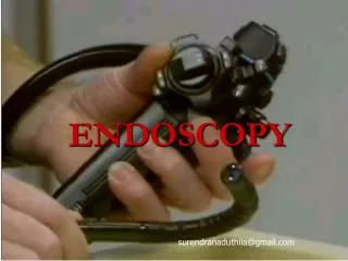
192 8-Principles of Endoscopy. an endoscope, light source, and irrigating fluid irrigation fluids include sterile water, glycine , or normal saline. If electrocautery use is anticipated, a solution free of electrolytes should be used.

E N D
192 8-Principles of Endoscopy • an endoscope, light source, and irrigating fluid irrigation fluids include sterile water, glycine, or normal saline. If electrocautery use is anticipated, a solution free • of electrolytes should be used
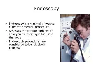
Video-endoscopic units, comprising a light source, camera for the endoscope, image processor and recorder, and monitor, are usually arranged on a mobile tower, are transmitted to the image processor by a camera attached to the eyepiece and displayed on a viewing monitor
Cystourethroscopy is used to directly visualize the anterior urethra, posterior urethra, and • the bladder. of the most common indications for cystourethroscopy is the evaluation of microscopic and gross hematuria. Other indications for cystourethroscopy include evaluation of voiding symptoms, surveillance of urothelial carcinoma, foreign body removal, and assisting in difficult placement • of a catheter.
Endoscope sizes are expressed using the French (Fr) scale and refer to the outer circumference in millimeters. Pediatric endoscopes are generally 8 to 12 Fr whereas adult scopes range from 16 to 25 Fr. The size of the endoscope selected will depend on the specific procedure performed, the need for additional working instruments, and the degree of irrigant flow that will be required, but in general the smallest diameter endoscope that will accomplish the goals of the procedure is selected to minimize genitourinary tract trauma.
UPPER URINARY TRACT ENDOSCOPY • Indications • Ureteroscopyis a standard urologic technique that provides direct visualization of the upper urinary tract, facilitating both diagnostic and therapeutic interventions • Ureteroscopy is most • commonly used for the treatment of nephrolithiasis
Ancillary Equipment • Wires. Guidewires used during retrograde instrumentation • serve to provide access to a particular area of the urinary tract and • serve as a guide to pass catheters, stents, and sheaths . • Guidewire properties vary with • respect to length, diameter, composition, tip design, surface • coating, and shaft rigidity. Guidewire diameters and lengths range • from 0.018 to 0.038 inch and 145 to 280 cm, respectively. • The • ideal guidewire should have a flexible lubricous tip allowing for • easy atraumatic passage through a tortuous, obstructed ureter • while providing sufficient rigidity of the shaft for the passage of • catheters and instruments.
Care should be taken to use only as much irrigation needed to provide a clear visual field. • Utilizing the minimum amount of irrigation necessary to • provide a clear view during ureteroscopy minimizes stone • migration, bleeding from hydrodistention, and pyelolymphatic • or pyelovenous backflow. l The holmium:YAG laser is the gold standard for ureteroscopic intracorporeal lithotripsy.
Complications of ureteroscopic • basketing range widely in severity and include ureteral • avulsion, intussusception, abrasion, perforation, postoperative • stricture formation, and basket breakage or • entrapment
PREPARATION FOR SURGERY • Patient Factors That Increase the Risk of Infection • Advanced age • Anatomic anomalies • Poor nutritional status • Smoking • Chronic corticosteroid use • Immunodeficiency • Chronic indwelling hardware • Infected endogenous/exogenous material • Distant coexistent infection • Prolonged hospitalization
Skin Preparation Sterile skin preparation is fundamental in the prevention of SSI for any procedure. Currently, the most commonly used skin antiseptics are alcohol, povidone-iodine, or chlorhexidine based. Whichever antiseptic is chosen, the solution should be applied in concentric circles from the center of the surgical site and be allowed to dry before incision. A recent review from the did not find sufficient evidence to recommend one skin preparation over another . Furthermore, although the CDC clearly recommends preoperative showering/bathing to reduce SSI, there is no evidence that bathing with an antiseptic solution reduces the rate of infection. Regarding hair removal, the CDC recommends that if hair removal is performed, it should be performed immediately before the surgical procedure and performed with clippers (rather than shaving .
PATIENT ENVIRONMENT • Patient Temperature There are two primary reasons for hypothermia to develop in the operating room. Anesthetic agents induce peripheral vasodilation redistributing heat from the core (trunk, head) with resultant drop in immediate core temperature after induction. Throughout the rest of the procedure, radiation and conductive heat loss account for most of the heat loss during a surgical procedure. Normothermia is defined as core temperature between 36° C and 38° C, and even hypothermia of 1° C to 2° C results in adverse effect. mild hypothermia (decrease of 1°C) resulted in a 16% increase in estimated blood loss and 22% increase in transfusion requirements
The increased bleeding risk is thought to result from a hypothermia-associated decrease in clotting cascade enzymatic function and platelet aggregation. Even more significant is the increase in the risk of surgical site infections (SSI) associated with mild hypothermia (34° C to 36° C). Hypothermia was associated with a three times increased risk of wound infection and a 2.6-day increase in hospitalization.
Strategies to improvement maintenance of :normothermia including regular use of warming blankets, warmed intravenous fluids, warmed irrigation fluids (especially during TURP and other prolonged endoscopic procedures), warmed/humidified CO2 gas during laparoscopy, and increase in ambient operating room temperature.
Skin Preparation Sterile skin preparation is fundamental in the prevention of SSI for any procedure. Currently, the most commonly used skin antiseptics are alcohol, povidone-iodine, or chlorhexidine based. Whichever antiseptic is chosen, the solution should be applied in concentric circles from the center of the surgical site and be allowed to dry before incision. A recent review from the Cochrane database did not find sufficient evidence to recommend one skin preparation over another . there is no evidence that bathing with an antiseptic solution reduces the rate of infection. Regarding hair removal, the CDC recommends that if hair removal is performed, it should be performed immediately before the surgical procedure and performed with clippers (rather than shaving) (Mangram et al, 1999)
Patient Safety : Three causes of immediately preventable injuries are retractor-associated injuries, thermal injuries and patient position–related injuries. 1-increased rate of neuropathy (especially femoral nerve) following laparotomy with self-retaining retractors versus without self-retaining retractors . Careful attention to be certain that the lateral blades do not directly compress the psoas muscle and only cradle the rectus abdominal muscles will ensure avoidance of femoral neuropathy
2-In both endoscopic and laparoscopic surgery, high-wattage light sources are used to illuminate the operative field. While illuminated, the ends of the light cords can result in burns when in direct contact with the patient (even through draping). These light sources should be turned off at all times when not in use.