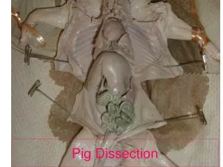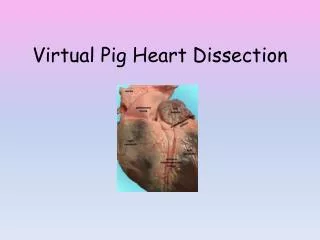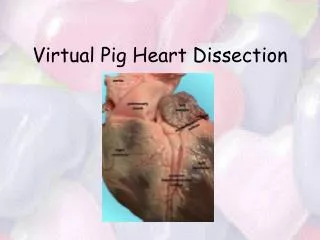
Pig Heart Dissection: A Comprehensive Guide to Anatomical Exploration
Dive into the fascinating world of anatomy with our Pig Heart Dissection guide. Learn how to properly prepare for dissection using essential surgical supplies like scalpels, scissors, and hemostats. Explore the exterior and interior anatomy of the heart, identifying key structures such as the auricles, coronary vessels, and heart valves. Follow step-by-step instructions to locate the great vessels, understand blood flow pathways, and familiarize yourself with important anatomical landmarks. Don't forget your gloves, and remember to tidy up after your exploration!

Pig Heart Dissection: A Comprehensive Guide to Anatomical Exploration
E N D
Presentation Transcript
Dissection Tray & Pig Heart Don’t forget your gloves!
Slide blade on scalpel handle. Peel back blade packet halfway. Slide over narrow tip of scalpel handle.
Base Apex
Auricles Ear-shaped extensions of theatria. Rt. Lt.
The auricles wrap around to the anterior side of the heart. Anterior
The Coronary Vessels The cardiac muscle receives oxygen for metabolism and releases waste products into coronary vessels.
The Great Vessels The large veins & arteries, which transport blood to and from the heart muscle.
Anterior View--Aortic Arch Aorta Arising from the left ventricle of the heart is the largest artery of the body, the aorta. Rt. Lt.
Anterior View--Aortic Arch Lt. common carotid Brachiocephalic trunk (innominate artery) Rt. Lt.
Aorta: Posterior View Aorta Lt. Rt.
Posterior View--Vena Cavae Superior Vena Cava Inferior Vena Cava Lt. Rt.
Posterior View--Pulmonary Artery Lt. Pulmonary Artery Lt. Rt.
Posterior View--Pulmonary Veins Rt. Lt. Pulmonary veins are inferior to pulmonary arteries.
Rt. Pulmonary Vein Lt. Pulmonary Vein
Open heart on dissection tray. Anterior half Posterior half Rt. Lt.
Rt. Atrium Lt. Atrium aorta Septum Rt. Lt.
aorta Width of right ventricular wall Width of left ventricular wall Septum Lt. Ventricle Rt. Ventricle
Tricuspid valve Cusp of valve Septum Papillary muscles Chordaetendinae
Mitral valve Septum
cusp Septum
Chordae tendinae Chordae tendinae
Check points--Exterior Heart Students should be able to point out: • Apex & Base of heart • Coronal Plane • Anatomical Right & Left Sides of heart • Anterior & Posterior Sides of heart • Auricles • Coronary Vessels
Check points--Interior Heart Students should be able to point out: • Septum • Atria & Ventricles • Tricuspid Valve, Pulmonary Valve • MitralValve, Aortic Valve • Chordae Tendinae • Papillary Muscles
Final Check point Students should be able to trace pathway of blood through great vessels and heart. (begin with vena cava and end with vena cava)
That’s All folks! …and don’t forget to clean up.
Borrowed from the South Carolina Dept of Education: http://ed.sc.gov/agency/Standards-and-Learning/Career-and-Technology-Education/old/cate/health_sciences/hsteteacherresourceguide/PigHeartDissection.ppt