
SlideServe
Menu
Préférences
© SlideServe · v2026

Analysis of LmPDI Domain Structure and Amino Acid Alignment Among Leishmania Species

wendi avatar
1 diapositives · 173 Views · Sep 13, 2014

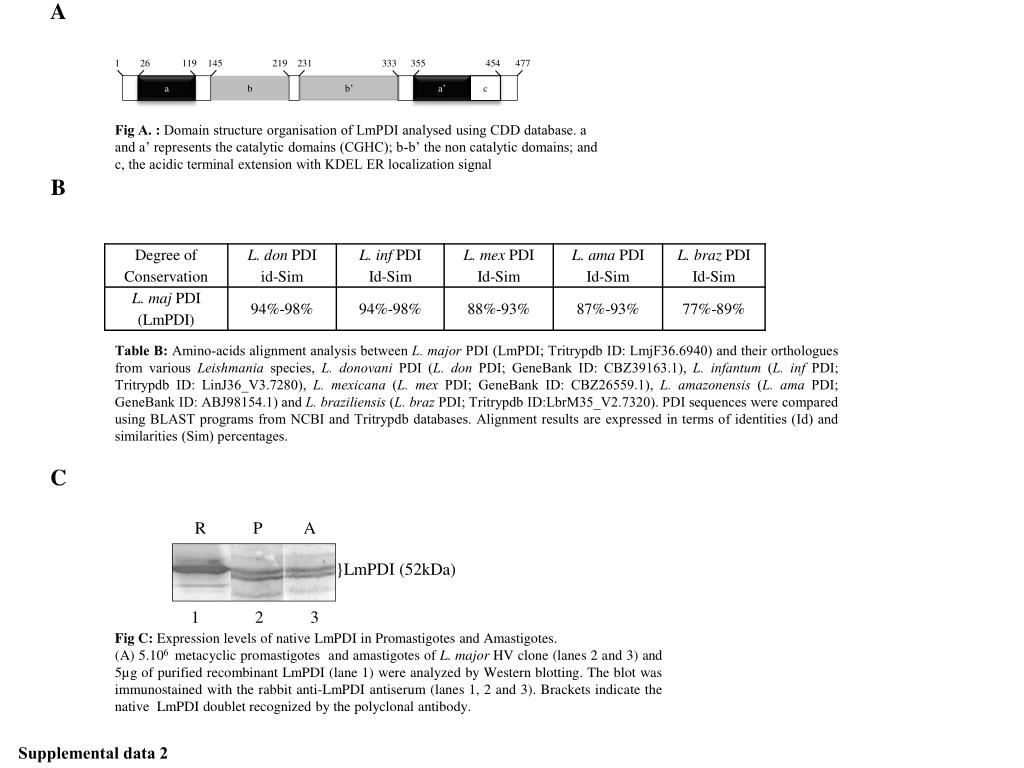
This study investigates the domain structure of LmPDI using the CDD database, identifying catalytic (CGHC) and non-catalytic domains, along with an acidic terminal extension featuring a KDEL ER localization signal. Amino acid alignments between LmPDI and its orthologues across different Leishmania species were performed using BLAST tools, providing insights into sequence identities and similarities. Additionally, expression levels of native LmPDI in both promastigotes and amastigotes were assessed through Western blotting, revealing important data about its recognition by specific antibodies.
Analysis of LmPDI Domain Structure and Amino Acid Alignment Among Leishmania Species - slide 1
01 / 1Analysis of LmPDI Domain Structure and Amino Acid Alignment Among Leishmania Species
1 / 17

Télécharger la présentation

Analysis of LmPDI Domain Structure and Amino Acid Alignment Among Leishmania Species
Une image / un lien ci-dessous est fourni (tel quel) pour télécharger la présentation

Politique de téléchargement : le contenu du site vous est fourni TEL QUEL pour votre information et votre usage personnel ; il ne peut être vendu, concédé sous licence ni partagé sur d’autres sites sans l’accord de son auteur.Le contenu vous est fourni TEL QUEL, pour votre information et votre usage personnel uniquement. Téléchargez la présentation en cliquant sur ce lien. Lors du téléchargement, si vous ne parvenez pas à télécharger une présentation, l’éditeur a peut-être supprimé le fichier de son serveur.Pendant le téléchargement, si vous ne parvenez pas à obtenir une présentation, le fichier a peut-être été supprimé par l’éditeur.

F I N

Plus de contenu associé

Transcription de la présentation
  1. A Fig A. : Domain structure organisation of LmPDI analysed using CDD database. a and a’ represents the catalytic domains (CGHC); b-b’ the non catalytic domains; and c, the acidic terminal extension with KDEL ER localization signal B 1 26 119 145 219 231 333 355 454 477 a b b’ a’ c Table B: Amino-acids alignment analysis between L. major PDI (LmPDI; Tritrypdb ID: LmjF36.6940) and their orthologues from various Leishmania species, L. donovani PDI (L. don PDI; GeneBank ID: CBZ39163.1), L. infantum (L. inf PDI; Tritrypdb ID: LinJ36_V3.7280), L. mexicana (L. mex PDI; GeneBank ID: CBZ26559.1), L. amazonensis (L. ama PDI; GeneBank ID: ABJ98154.1) and L. braziliensis (L. braz PDI; Tritrypdb ID:LbrM35_V2.7320). PDI sequences were compared using BLAST programs from NCBI and Tritrypdb databases. Alignment results are expressed in terms of identities (Id) and similarities (Sim) percentages. C R P A }LmPDI (52kDa) 1 2 3 Fig C: Expression levels of native LmPDI in Promastigotes and Amastigotes. (A) 5.106 metacyclic promastigotes and amastigotes of L. major HV clone (lanes 2 and 3) and 5µg of purified recombinant LmPDI (lane 1) were analyzed by Western blotting. The blot was immunostained with the rabbit anti-LmPDI antiserum (lanes 1, 2 and 3). Brackets indicate the native LmPDI doublet recognized by the polyclonal antibody. Supplemental data 2