Download
1 / 6
60 likes | 186 Vues
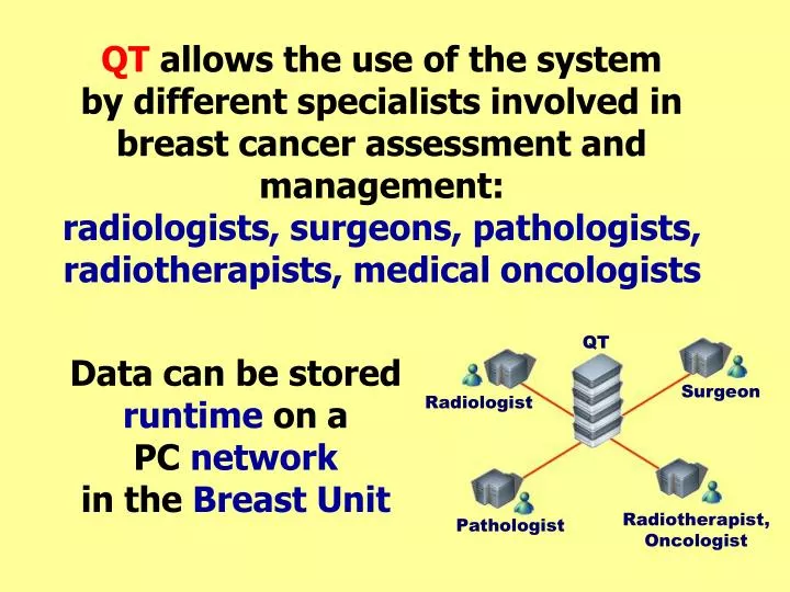
QT allows the use of the system by different specialists involved in breast cancer assessment and management: radiologists, surgeons, pathologists, radiotherapists, medical oncologists. QT. Data can be stored runtime on a PC network in the Breast Unit. Surgeon. Radiologist.

E N D
QT allows the use of the system by different specialists involved in breast cancer assessment and management: radiologists, surgeons, pathologists, radiotherapists, medical oncologists QT Data can be stored runtime on a PC network in the Breast Unit Surgeon Radiologist Radiotherapist, Oncologist Pathologist
QT includes: 22 data-entry forms 25 Outcome measures 5 automatic reports (History, Assessment, Surgery) EpiInfo 6 Emulator (for complex statistical analysis)
Guided data-entry: colors help to distinguish minimum set fields (white) from other less important items (green). Not pertinent fields are grey
Outcome measures easily calculated with just one click
First Screening CentreS.John HospitalBourbon Street, 31LT160, Caribbean Borough Date 06/11/2000 Mrs. Jenny Smith Diagnostic report Screening mammogram carried out on 02/08/2001.Assessment session carried out on 14/08/2001. Palpable lesion of 15 mm. measured by ultrasound located in inferior-internal area of right breast. At mammography is an abnormality of indeterminate significance (R3)(06/08/2001). Microcalcifications state is unknown. Ultrasound result is lesion presenting malignant features (U5) (14/08/2001). FNA has not been performed. Core biopsy guided by ultrasound has been performed (15/08/2001) with the following hystological result: "B5-Malignant". Recommendation (15/08/2001): exeresis. The following axillary operation was performed: sentinel lymphnode. Histopathological diagnosis: Invasive carcinoma. • Automatic reports • available: • Diagnostic rep. • Surgical rep. • Histological rep. • History rep. • Surgery description rep.
Multidimensional analysis up to 4 variables and graphical reports