Mandible BY: DR.Yahya Alfarra
190 likes | 512 Vues
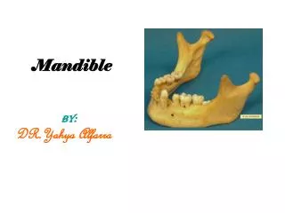
Mandible BY: DR.Yahya Alfarra. Mandible. Mandible consists of the body & 2 rami The two halves of body of Md. Meet each other in the median plane at symphysis menti . The lower border of the body of Md. Meets post. Border of the ramus at angle of Md. Which change by age.

Mandible BY: DR.Yahya Alfarra
E N D
Presentation Transcript
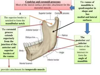
Mandible • Mandible consists of the body & 2 rami • The two halves of body of Md. Meet each other in the median plane at symphysis menti. • The lower border of the body of Md. Meets post. Border of the ramus at angle of Md. Which change by age
symphysis menti from mental protuberance as triangualr elevation having mental tubercle on each side. • Lateral to symphysis menti there is mental foramen which transmit mental N. & Vesseles • thick ridge called oblique line.
Inside the body of Md. There is Md. Canal , the entrance of this canal lies on inner surface of ramus & is called Mandibualr foramen.
Upper border of body of Md. Termed alveolar process which carries the socket or alveoli of the teeth. • Lower border of body of Md. Termed base of Md. That gives attachment with …….. Ms .
The ramus of Md. Has 2 process projecting from upper border: The condylar process post. Coronoid process ant. • The condylar process comprise head & neck of Md. • Md. Notch lies between 2 process Note: • The front neck of Md. Give insertion to lateral pterygoid Ms. • The coronoid process give insertion to temporalis Ms. • The lateral surface of ramus of Md. Give insertion to masseter Ms. & it’s cover by parotid gland .
The medial border of foramen is projecting to form lingula which sphenomandibualr ligament attachment, • just behind lingula, there is mylohyoid groove that lodges mylohyoid N.& V.
Posterior to the third molar a triangular shallow fossa is outlined; it is called the retromolar triangle.