Download
1 / 12
130 likes | 228 Vues
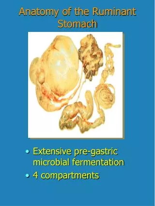
Anatomy of Stomach and Duodenum. Blood Supply. Lymphatic Drainage. Lymphatic drainage Lymph nodes draining the stomach are numbered and divided into 4 levels, as follows:

E N D
Lymphatic Drainage Lymphatic drainage Lymph nodes draining the stomach are numbered and divided into 4 levels, as follows: Level I (perigastric lymph nodes) - Right paracardiac (1), left paracardiac (2), along lesser curvature (3) along greater curvature (4), suprapyloric (5), infrapyloric (6) Level 2 - Along LGA (7), along CHA (8), along celiac axis (9), at splenic hilum (10), along splenic artery (11) Level 3 - In hepato-duodenal ligament (12), behind duodenum and pancreas head (13), at the root of small bowel mesentery (14) Level 4 - Mesocolic (15), paraaortic (16)
Nerves • Intrinsic- Myenteric plexus of Auerbach and Meissner’ssubmucosal plexus. • Extrinsic- right ( posterior) and left (anterior) vagus nerve. Nerves of Latarjet( crow’s foot) • Sympathetic- caeliac ganglion
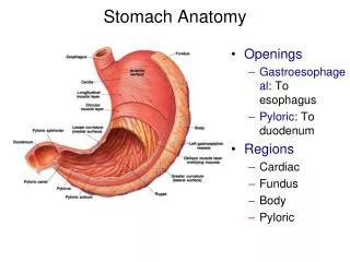
Microscopy • Mucous cells- pylorus and duodenum • Parietal cells- gastric crypts in the body. Secrete acid. • Chief cells- gastric crypts. Secrete pepsinogen. • Endocrine- G cells in the antrum: gastrin. Enterochromaffin like cells in the body: histamine. D cells: somatostatin. Peptide and neuropeptides. • Duodenum- mucous cells, endocrine cells- secretin-cck)
Gastric acid secretion • Cephalic phase- vagus • Gastric phase- mainly gastrin also vagus • Intestinal phase- secretin, neuropeptides, somatostatin
Gastric mucosal barrier • Integrity of gastric mucosa is due to viscid layer of mucopolysaccharide produced by mucous cells lining stomach • Buffering capacity is enhanced by bicarbonate ions. • Barrier is broken by bile, shock, alcohol, NSAID, steroids.
Investigations • Flexible gastroduodenoscopy • Ultrasonography-conventional, endoluminal • CT scan • MRI • PET- ST scan • Laparoscopy • Gastric emptying studies- motility disorders, post-op • Angiography • Barium meal.
Helicobacter Pylori • Described by Bircher in 1874 • Warren and Marshall demonstrated that it causes gastritis • Spiral shaped, detected on gastric biopsy by Giemsa stain, EthinStarey silver stain. • 13C and 14C Breath test and CLO test on gasric biopsy • Spreads by faeco-oral route. • Causes chronic gastritis, peptic ulcers, gastric cancers.
It can hydrolyseure to produce ammonia which by negative feed back releases gastrin stimulating acid secretion • Treatment- PPI + metronidazole+ amoxycillin/ clarithromycine. For 2 weeks. Re infections are common