
RESPIRATORY SYSTEM
Explore the structures, mechanics, and functions of the respiratory system, including gas transfer, regulation of blood pH, and pumping action. Learn about the upper and lower respiratory tracts, as well as the functions of the nose and pharynx.

RESPIRATORY SYSTEM
E N D
Presentation Transcript
CONTENTS:- • Introduction,generalorganisation • Structures of respiratory system • Mechanics of respiration, compliance • Lung volumes and capacities • Pulmonary and alveolar ventilation • Physical principles of gaseous exchange • Transport of respiratory gases • Control of respiration • Pulmonary function tests.
INTRODUCTION Respiratory system constitutes one of the important system as it contributes the vital provision of supply of oxygen to the body, where cells continuously use oxygen for metabolic reactions which in turn releases energy. This energy is from the nutrient molecules which provide ATP.
DEFINITION:- Respiration is the process by which oxygen from the lungs is carried by the blood to the tissues and carbon dioxide formed in the tissues by metabolic activity is carried by the blood to the lungs and is expired outThe PROCESS OF RESPIRATION involves four stages:-VENTILATION:-Ventilation means the passage of air in and out of lungs during Inspiration and expiration respectively .
INTRAPULMONARYGAS MIXING:-Intrapulmonary gas mixing or distribution of oxygen rich inspired air with the air already present in the lungs.DIFFUSION:- Diffusion which means gas transfer across the alveolar capillary membrane due to tension gradient.PERFUSION:- Perfusion means flow of adequate quantity of blood through the lungs so that the diffused gases are carried away.
FUNCTIONS OF RESPIRATION • GAS TRANSFER: • Transfer of O2 from the alveoli to the venous blood and CO2 in the opposite direction. • REGULATION OF pCO2 OF BLOOD: • The most important function of respiration is to keep the arterial pCO2 at 40 mmHg which is essential for many vital functions of the body. • REGULATION OF PH OF BLOOD: • By the reversible reaction, it maintains the pH of blood • H2CO3H+ + HCO3-
EXCRETION OF CERTAIN VOLATILE GASES: • For example- chloroform, ether, ammonia, etc. • PUMPING ACTION: • The rhythmic movement of the diaphragm and the chest wall causes rhythmic alteration of pressure in the abdomen and chest cavity. This assists in drawing blood from the lower part of the body to the abdomen and then to chest and thus helps in maintaining venous inflow to the heart.
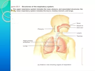
RESPIRATORY TRACT • The whole respiratory tract is divided in two parts :- • UPPER RESPIRATORY TRACT- • It extends from the nasal cavity to the vocal cords i.e. (nose, pharynx and associated structures) • LOWER RESPIRATORY TRACT- • It extends from the vocal cord to the alveoli i.e (larynx, trachea, bronchi, lungs.)
STRUCTURES OF THE RESPIRATORY TRACT • CONTENTS- • Nose • Pharynx • Larynx • Trachea • Bronchi • Lungs
NOSE AND NASAL CAVITY • CONTENTS • POSITION AND STRUCTURE • LINING OF THE NOSE • FUNCTIONS OF THE NOSE- • (A) RESPIRATORY FUNCTIONS OF THE NOSE • (B) OLFACTORY FUNCTIONS OF THE NOSE
POSITION AND STRUCTURE :- • The nasal cavity is the main route of air entry , and consists of a large irregular cavity divided in to two equal passages by a septum . The posterior bony part of the septum is formed by the perpendicular plate of the ethmoid bone and vomer. Anteriorly, it consists of hyaline cartilage . • LINING OF THE NOSE :- • The nose is lined with very vascular ciliated columnar epithelium which contains mucus secreting goblet cells. At the anterior nares this blends with the skin and posterioly it extends in to the nasal part of the pharynx.
FUNCTIONS OF THE NOSE • RESPIRATORY FUNCTONS OF THE NOSE:- • WARMING:- This is due to the immense vascularity of the mucosa. This explains the large blood loss when a nosebleed occurs . • 2) FILTERING AND CLEANING :- This occurs as hairs at the anterior nares trap large particles. Smaller particales such as dust and microbes settle and adhere to the mucus. Mucus protects the underlying epithelium from irritation and prevents drying. Synchronous beating of the cilia wafts the mucus towards the throat where it is swallowed or coughed up.
3) HUMIDIFICATION:- As air travels over the moist mucosa , it becomes saturated with water vapour . irritation of the nasal mucosa results in sneezing, a reflex action that forcibly expels an irritant. IV) OLFACTORY FUNCTIONS OF THE NOSE :- The nose is the organ of the sense of smell . Nerve endings that detect smell are located in the roof of the nose in the area of the cribriform plate of the ethmoid bones and the superior conchae. These nerve endings are stimulated by airborne odours. The resultant nerve impulses are conveyed by the olfactory nerves to the brain where the sensation of smell is perceived.
PHARYNX • CONTENTS:- • POSITION OF PHARYNX • DIVISION OF PHARYNX • BLOOD AND NERVE SUPPLY • FUNCTIONS OF PHARYNX
1. POSITION OF PHARYNX:- • The pharynx is a tube 12 to14 cm long that extends from the base of the skull to the level of the 6th cervical vertebra.it lies behind the nose mouth and larynx and is wider at its upper end • 2. DIVISION OF PHARYNX:- • (A)THE NASOPHARYNX :- • The nasal part of the pharynx lies behind the Nose Above the level of the soft palate • (B) THE OROPHARYNX:- • The oral part of the pharynx lies behind the Mouth extending from below the level of the soft palate to the level of the upper part of the body of the third cervical vertebra
(C) THE LARYNGOPHARYNX :- The laryngeal part of the pharynx extends From the oropharynx above the continues as the oesophagus below from the level of the 3rd to the 6th cervical vertebrae . 3. BLOOD AND NERVE SUPPLY :- Blood supply is by the facial artery . Nerve supply is by the PARASYMPATHETIC by the :- Vagus nerve and glosso pharyngeal nerve. SYMPATHETIC by the :- Superior cervical ganglia.
4. FUNCTIONS OF THE PHARYNX:- I) WARMING AND HUMIDIFYING :- By the same methods as in the nose the air is further warmed and moistened as it passes through the pharynx. II) PASSAGE WAY FOR AIR AND FOOD:- Air passes through the nasal And oral section and food Through the oral and laryngeal section.
LARYNX • CONTENTS- • POSITION • BLOOD AND NERVE SUPPLY • FUNCTIONS
POSITION :- The larynx or voice box extends from the root of the tongue and the hyoid bone to the trachea. It lies infront of the laryngo pharynx at the Level of the 3rd,4th,5th and 6th cervical vertebra BLOOD AND NERVE SUPPLY :- • BLOOD SUPPLY:- By the superior and inferior laryngeal arteries. • NERVE SUPPLY :- • a) PARASYMPATHETICS :- By the superior laryngeal And recurrent Laryngeal nerves. • b) SYMPATHETICS:-Superior cervical ganglia,
FUNCTIONS :- • SPEECH:- • This occurs during expiration when the sounds produced by the vocal cords are manipulated by the tongue,cheek and lips. • PROTECTION OF THE LOWER RESPIRATORY TRACT:- • During Deglutition the larynx move upwards occulding the opening in to it from the pharynx and the hinged epiglottis closes over the larynx this ensure that food passes in to the oesophagus and not into the lower respiratory tract. • PASSAGEWAY FOR AIR:- • This is between the pharynx and trachea
HUMIDIFYING FILTERING AND WARMING :- • This processes continue As inspired air travel Through the larynx.
TRACHEA CONTENTS:- • POSITION • STRUCTURE • BLOOD AND NERVE SUPPLY • FUNCTION
POSITION:- The trachea or wind pipe is a continuation of the larynx and Extends downwards to about the level of the 5th thoracic Vertebra. STRUCTURE :- The trachea is a cartilaginous membranous tube about 10 or 11cm. long it is not quite cylindrical being flattened posteriorly. Its external diameter from side to side is about 2cm in the adult male and 1.5 in the adult female it is kept patent by incomplete C-shaped rings of cartilage on its anterio lateral wall which keeps air tubes open.
BLOOD AND NERVE SUPPLY :- BLOOD SUPPLY :-By the inferior thyroid and bronchial arteries. NERVE SUPPLY:- a)PARASYMPATHETICS:-By the recurrent laryngeal nerve b)SYMPATHETICS:- By the ganglia
FUNCTIONS:- • SUPPORT AND PATENCY :-trachea is kept patent by incomplete C-shaped ring Of cartilage on its anterotateral wall which keeps • Air tube open. • COUGH REFLEX :-Cough is protective reflex by means of which respiratory Passage are kept free form foreign matter. • WARMING,HUMIDIFYING AND FILTERING :- This continues as the nose, Although air is normally Saturated and at body temperature when it reaches the trachea.
APPLIED PHYSIOLOGY:- TRACHEOSTOMY AND INTUBATION Several condition may Block air flow by Obstructing the trachea FOR EXAMPLE :-The rings of cartilage that support the trachea may collapse due to a crushing injury to the chest inflammation of the mucous membrane may cause it to Swell so much that the airway closes or vomit or a foreign object may be aspirated. Methods are used to reestablish air flow past a tracheal obstruction if the obstruction is superior to the level of the larynx tracheostomy an operation to make an opening in to the trachea may be performed.
BRONCHI CONTENTS:- • STRUCTURE • BLOOD AND NERVE SUPPLY • FUNCTIONS
STRUCTURE :- The bronchi is progressively subdivided in to bronchioles, Terminal bronchioles, respiratory bronchioles, albeolar ducts and finally alveoli. Situated from the superior border of the 5th thorasic vertebra divided in to a right primary bronchus which goes in to the right lungs and a left primary bronchus which goes in to the left lungs. BLOOD AND NERVE SUPPLY:- BLOOD SUPPLY :- By the right & left Bronchial arteries NERVE SUPPLY :-PARASYMPATHETICS & SYMPATHETIC NERVE
3) FUNCTIONS :- • CONTROL OF AIR ENTRY :- The diameter of the respiratory passages is Altered by contraction and relaxation of the Involuntary muscles in their walls, thus regulating the volume of entering the Lungs. These changes are controlled by the autonomic nerve supply parasympathetics causes constriction and sympathetics stimulation dilatation . • WARMING AND HUMIDIFYING • SUPPORT AND PATENCY • REMOVAL 0F PARTICULATE MATTER • COUGH REFLEX.
LUNGS CONTENTS:- • INTRODUCTION • POSITION AND STRUCTURE • COVERING OF LUNGS • LOBES, FISSURES AND LOBULES • ALVEOLI • SURFACTANT • RESPIRATORY MEMBRANE • BLOOD SUPPLY • NERVE SUPPLY • APPLIED PHYSIOLOGY
INTRODUCTION :- The lungs (=lightweights, because they float), are paired cone- shaped organs in the thoracic cavity. They are separated from each other by the heart and other structures in the mediastinum while thoracic Cavity & mediastinum form two different chambers for two lungs.
POSITION AND STRUCTURE The lungs extend from the diaphragm to just slightly superior to the clavicles and lie against the ribs anteriorly and posteriorly. The broad inferior portion of the lung, the base, is concave and fits over the convex area of the diaphragm. The narrow superior portion of the lungs is the apex. The surface of the lungs lying against the ribs, called the costal surface, matches the rounded curvature of the ribs. Medially, the left lung also contains a concavity, the cardiac notch, in which the heart lies
The mediastinal (medial) surface of each lung contains the region, the hilus, through which bronchi, pulmonary blood vessels, lymphatic vessels, and nerves enter and exit. These structures are held together by pleura and connective tissue and constitute the root of lung. Due to the space occupied by the heart, the left lung is about 10% smaller than the right lung.
COVERING OF LUNGS- Two layers of serous membrane collectively called the pleural membrane, enclose and protect each lung. The superficial layer lines the wall of the thoracic cavity and is called the parietal pleura, the deep layer, the visceral pleura, covers the lungs themselves . Between the visceral and parietal pleurae is a small space, the pleural cavity.
Pleural cavity contains a small amount of lubricating fluid secreted by the membranes. This fluid reduces friction between the membranes, allowing them to slide easily over one another during breathing. Pleural fluid also causes the two membranes to adhere to one another just as a film of water causes two slides to stick together , a phenomenon called surface tension.
LOBES, FISSURES AND LOBULES:- The right lung is divided into three lobes-superior, middle and inferior. The left lung is smaller and is divided into two lobes-superior and inferior. Each lung is divided into lobes by one or more fissures .Both the lungs have oblique fissures . Right lung also has horizontal fissure. The oblique fissure in the left lung separates the superior lobe from the inferior lobe
Each lobe receives its own secondary (lobar)bronchus .Thus, the right primary bronchus gives rise to three secondary bronchi called the superior, middle and inferior secondary bronchi, whereas the left primary bronchus gives rise to superior and inferior secondary bronchi. Within the substance of lung, the secondary bronchi give rise to the tertiary bronchi. The segment of lung tissue that each tertiary bronchus supplies is called a “Broncho pulmonary segment” .
ALVEOLI Around the circumference of the alveolar ducts are numerous alveoli and alveolar sacs. An alveolus is a cup-shaped outpouching lined by simple squamous epithelium and supported by a thin elastic basement membrane. An alveolar sac consists of two or more alveoli that share a common opening.
The walls of alveoli contains two types of alveolar epithelial cells- 1.)Type I alveolar cells. 2.)Type II alveolar cells. The Type I alveolar cells, the predominant cells, are simple squamous epithelial cells that form a nearly continuous lining of the alveolar wall. The Type II alveolar cells, also called septal cells, are fewer in number and are found in between type I alveolar cells.
FUNCTIONS OF ALVEOLAR CELLS:- • Type I alveolar cells are main sites of gaseous exchange. • Type II alveolar cells, which are rounded or cuboidal epithelial cells whose free surfaces contain microvilli, secrete alveolar fluid, which keeps the surface between cells and air moist. Included in the alveolar fluid is SURFACTANT. • Asssociated with alveolar walls are alveolar macrophages (dust cells), wandering phagocytes that remove the fine dust particles and other debris in the alveolar spaces.
SURFACTANT- DEFINATION- Any surface acting material or agent that is responsible for lowering surface tension of fluid is called surfactant. The surfactant present in the alveoli of lungs is called as pulmonary surfactant. It is a complex detergent like mixture of phospholipids and lipoproteins. It is secreted by Type II alveolar epithelial cells.
FUNCTIONS OF SURFACTANT- • They reduces surface tension in alveoli of lungs & prevents collapsing tendency of lungs. • Surfactant is responsible for stabilization of alveoli which have tendency to deflate. • It plays an important role in inflation of lungs during birth.
RESPIRATORY MEMBRANE- The exchange of oxygen & carbon dioxide between the air spaces in the lungs and the blood capillaries takes place by diffusion across the alveolar and capillary walls, which together form the respiratory membrane. Extending from the alveolar airspace to blood plasma, the respiratory membrane consists of four layers:-


