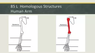
Arm Muscle and Structures
130 likes | 169 Vues
Explore the muscles that move the forearm and hand with detailed illustrations, including triceps brachii, biceps brachii, and more. Learn about bone structures and ligaments in the arm for enhanced understanding.

Arm Muscle and Structures
E N D
Presentation Transcript
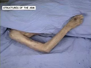
Arm Muscle and Structures Blank Pics
Figure 11-16a Muscles That Move the Forearm and Hand (Part 2 of 2) Triceps brachii, long head Triceps brachii, lateral head Brachioradialis Olecranon of ulna Anconeus Extensor carpi radialis longus Flexor carpi ulnaris Extensor carpi radialis brevis Extensor carpi ulnaris Abductor pollicis longus Extensor digitorum Extensor pollicis brevis Ulna Extensor retinaculum Posterior view, superficial layer
Figure 11-16a Muscles That Move the Forearm and Hand (Part 2 of 2)
Figure 11-16b Muscles That Move the Forearm and Hand (Part 2 of 2) Coracoid process of scapula Humerus Coracobrachialis Biceps brachii, short head Biceps brachii, long head Triceps brachii, long head Triceps brachii, medial head Brachialis Medial epicondyle of humerus Pronator teres Brachioradialis Flexor carpi radialis Palmaris longus Flexor carpi ulnaris Flexor digitorum superficialis Pronator quadratus Flexor retinaculum Anterior view, superficial layer
Figure 11-16b Muscles That Move the Forearm and Hand (Part 2 of 2)
Figure 8-4 The Right Humerus and Elbow Joint Greater tubercle Head Greater tubercle Lesser tubercle Intertubercular groove Anatomical neck Surgical neck Deltoid tuberosity Radial groove Shaft Olecranon fossa Radial fossa Coronoid fossa Lateral epicondyle Lateral epicondyle Medial epicondyle Capitulum Trochlea Trochlea Humerus Condyle Anterior surface Posterior surface Medial epicondyle Humerus Olecranon fossa Medial epicondyle Olecranon Trochlea Head of radius Trochlea of humerus Capitulum Ulna Coronoid process of ulna Head of radius Radial notch of ulna Elbow joint, anterior view Elbow joint, posterior view
Figure 8-5 The Right Radius and Ulna Olecranon Olecranon Trochlear notch Coronoid process Radial head Proximal radioulnar joint Radial notch Neck of radius Ulnar tuberosity Radial tuberosity ULNA ULNA ULNA RADIUS Lateral view of ulna, showing trochlear notch Interosseous membrane Ulnar notch of radius Distal radio-ulnar joint Ulnar head Ulnar head Styloid process of ulna Styloid process of radius Anterior view Posterior view
Figure 8-6a Bones of the Right Wrist and Hand RADIUS ULNA Lunate Scaphold Triquetrum Trapezium Pisiform Trapezoid I Capitate Hamate V IV Metacarpal bones III II Proximal phalanx Distal phalanx Anterior view
Figure 9-10 The Right Elbow Joint Showing Stabilizing Ligaments Tendon of bicepsbrachii muscle Annularligament Humerus Radialcollateralligament Humerus Antebrachialinterosseousmembrane Radialtuberosity Articularcapsule Antebrachialinterosseousmembrane Medialepicondyle Radius Ulnarcollateralligament Radius Ulna Ulna Olecranonof ulna Capitulum Annular ligament (coveringhead and neck of radius) Lateral view Medial view
Figure 9-10 The Right Elbow Joint Showing Stabilizing Ligaments