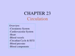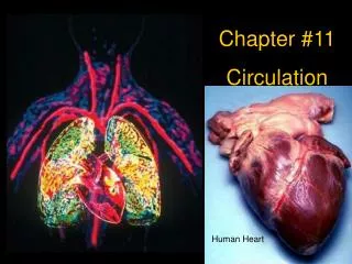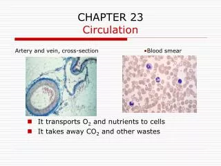
CHAPTER 23 Circulation
CHAPTER 23 Circulation. Modules 23.1 – 23.3. How Does Gravity Affect Blood Circulation?. As with all land animals, the giraffe and the corn snake are constantly subject to the force of gravity . The circulatory system keeps blood pumping despite gravity’s pull.

CHAPTER 23 Circulation
E N D
Presentation Transcript
CHAPTER 23Circulation Modules 23.1 – 23.3
How Does Gravity Affect Blood Circulation? • As with all land animals, the giraffe and the corn snake are constantly subject to the force of gravity
The circulatory system keeps blood pumping despite gravity’s pull • Muscle contractions help blood travel uphill in the veins of a giraffe’s long legs • The wriggling of the corn snake squeezes its veins and increases circulation
It transports O2 and nutrients to cells • It takes away CO2 and other wastes • Most animals have a circulatory system
23.1 The circulatory system associates intimately with all body tissues • Capillaries are microscopic blood vessels • They form an intricate network among the tissue cells Capillary Redbloodcell Figure 23.1A
No substance has to diffuse far to enter or leave a cell Capillary Diffusion ofmolecules INTERSTITIALFLUID Tissuecell Figure 23.1B
MECHANISMS OF INTERNAL TRANSPORT 23.2 Several types of internal transport have evolved in animals • In cnidarians and flatworms, the gastrovascular cavity functions in both • digestion • internal transport Mouth Circularcanal Figure 23.2A
Open systems • A heart pumps blood through open-ended vessels into spaces between cells • Most animals have a separate circulatory system, either open or closed Tubular heart Pores Figure 23.2B
A heart pumps blood through arteries and capillary beds • The blood returns to the heart via veins • Closed systems Capillary beds Arteriole Artery(O2-rich blood) Venule Vein Atrium Heart Artery(O2-poor blood) Ventricle Gillcapillaries Figure 23.2C
23.3 Vertebrate cardiovascular systems reflect evolution Gill capillaries • A fish has a single circuit of blood flow Heart: Ventricle (V) Atrium (A) Systemic capillaries Figure 23.3A
Lung capillaries • The pulmonary circuit • conveys blood between the heart and gas-exchange tissues • The systemic circuit • carries blood between the heart and the rest of the body • The cardiovascular system of land vertebrates has two circuits PULMONARYCIRCUIT A A V V Right Left SYSTEMICCIRCUIT Figure 23.3B Systemic capillaries
THE MAMMALIAN CARDIOVACULAR SYSTEM 23.4 The human heart and cardiovascular system typify those of mammals • The mammalian heart has two thin-walled atria that pump blood into the ventricles • The thick-walled ventricles pump blood to all other body organs
Pulmonaryartery Aorta Pulmonaryartery Superiorvena cava LEFTATRIUM RIGHTATRIUM Pulmonaryveins Pulmonaryveins Semilunarvalve Semilunarvalve Atrioventricularvalve Atrioventricularvalve Inferiorvena cava RIGHTVENTRICLE LEFTVENTRICLE Figure 23.4A
Superiorvena cava 7 Capillaries of Head and arms Pulmonaryartery Pulmonaryartery Capillariesof right lung Capillariesof left lung Aorta 9 6 2 3 3 4 11 Pulmonaryvein Pulmonaryvein 5 LEFT ATRIUM 1 RIGHT ATRIUM LEFT VENTRICLE RIGHT VENTRICLE 10 Aorta Inferiorvena cava Capillaries ofabdominal organsand legs 8 Figure 23.4B
23.5 The structure of blood vessels fits their functions • A single layer of epithelial cells forms capillary walls • Arteries and veins have smooth muscle and connective tissue • Valves in veins prevent the backflow of blood
Valve Epithelium Basementmembrane Epithelium Epithelium Smoothmuscle Smoothmuscle CAPILLARY Connectivetissue Connectivetissue ARTERY VEIN VENULE ARTERIOLE Figure 23.5
23.6 The heart contracts and relaxes rhythmically 1 Heart isrelaxed.AV valvesare open. 2 Atriacontract. • Diastole • Blood flows from the veins into the heart chambers • Systole • The atria briefly contract and fill the ventricles with blood • Then the ventricles contract and propel blood out SYSTOLE 0.1 sec 3 Ventriclescontract.Semilunarvalvesare open. 0.3 sec 0.4 sec DIASTOLE Figure 23.6
Cardiac output • The amount of blood pumped into the aorta by the left ventricle per minute • Heart valves prevent backflow
23.7 The pacemaker sets the tempo of the heartbeat • The SA node (pacemaker) generates electrical signals that trigger the contraction of the atria • The AV node then relays these signals to the ventricles Specializedmuscle fibers Pacemaker (SA node) AV node Rightatrium Rightventricle 1 2 3 4 ECG Figure 23.7
Control centers in the brain adjust heart rate to body needs • An electrocardiogram (ECG) is a recording of electrical changes in the skin resulting from the electrical signals in the heart
23.8 Connection: What is a heart attack? • A heart attack is damage that occurs when a coronary feeding the heart is blocked Aorta Rightcoronaryartery Leftcoronaryartery Blockage Dead muscle tissue Figure 23.8A
Blood vessel blockage is usually due to blood clots Connectivetissue Smoothmuscle Epithelium Plaque Figure 23.8B
23.9 Blood exerts pressure on vessel walls • Blood pressure depends on • cardiac output • resistance of vessels
Systolicpressure Diastolicpressure • Pressure is highest in the arteries • It drops to zero by the time the blood reaches the veins Relative sizes andnumbersof blood vessels Figure 23.9A
muscle contractions • breathing • one-way valves • Three factors keep blood moving back to the heart Direction ofblood flowin vein Valve (closed) Valve (open) Skeletal muscle Figure 23.9B
23.10 Connection: Measuring blood pressure can reveal cardiovascular problems • Blood pressure is measured as systolic and diastolic pressures Blood pressure120 systolic80 diastolic(to be measured) Pressurein cuffabove120 Pressurein cuffbelow120 Pressurein cuffbelow 80 Rubber cuffinflated with air Soundsaudible instethoscope Soundsstop Arteryclosed Artery 2 3 4 1 Figure 23.10
Hypertension is persistent systolic pressure higher than 140 mm Hg and/or diastolic pressure higher than 90 mm Hg • It is a serious cardiovascular problem
23.11 Smooth muscle controls the distribution of blood • Muscular constriction of arterioles and precapillary sphincters controls the flow through capillaries Precapillary sphincters Thoroughfarechannel Thoroughfarechannel Venule Arteriole Venule Arteriole Capillaries 2 1 Sphincters contracted Sphincters relaxed Figure 23.11
23.12 Capillaries allow the transfer of substances through their walls Figure 23.12A
leakage through clefts in the capillary walls • diffusion through the wall • blood pressure • osmotic pressure • The transfer of materials between the blood and interstitial fluid can occur by
Tissue cells Osmoticpressure Osmoticpressure Arterialend ofcapillary Venousend ofcapillary Bloodpressure Bloodpressure INTERSTITIALFLUID NET PRESSUREOUT NET PRESSUREIN Figure 23.12B
STRUCTURE AND FUNCTION OF BLOOD 23.13 Blood consists of cells suspended in plasma • Plasma is an aqueous solution of various substances
Withdrawblood Centrifuge Place in tube PLASMA 55% CONSTITUENT MAJOR FUNCTIONS CELLULAR ELEMENTS 45% Solvent forcarrying othersubstances CELL TYPE NUMBER(per mm3 of blood) FUNCTIONS Water Erythrocytes(red blood cells) Salts 5–6 million Transport ofoxygen (and carbon dioxide) Sodium Potassium Calcium Magnesium Chloride Bicarbonate Osmotic balance,pH buffering, andregulation ofmembranepermeability Leukocytes(white blood cells) Defense andimmunity 5,000–10,000 Plasma proteins Albumin Fibrinogen Immunoglobins(antibodies) Osmotic balance,pH buffering Clotting Immunity Lymphocyte Basophil Eosinophil Substances transported by blood Monocyte Nutrients (e.g., glucose, fatty acids, vitamins) Waste products of metabolism Respiratory gases (O2 and CO2) Hormones Neutrophil Platelets 250,000–400,000 Blood clotting Figure 23.13
23.14 Red blood cells transport oxygen • Red blood cells contain hemoglobin • Hemoglobin enables the transport of O2 Figure 23.14
23.15 White blood cells help defend the body • White blood cells function both inside and outside the circulatory system • They fight infections and cancer Basophil Eosinophil Monocyte Neutrophil Lymphocyte Figure 23.15
23.16 Blood clots plug leaks when blood vessels are injured • When a blood vessel is damaged, platelets respond • They help trigger the formation of an insoluble fibrin clot that plugs the leak Figure 23.16B
1 Injury to lining of bloodvessel exposes connectivetissue; platelets adhere 2 Platelet plug forms 3 Fibrin clot trapsblood cells Connectivetissue Plateletplug Platelet releases chemicalsthat make nearby platelets sticky Clotting factors from: Platelets Calcium andother factorsin blood plasma Damaged cells Prothrombin Thrombin Fibrinogen Fibrin Figure 23.16A
23.17 Connection: Stem cells offer a potential cure for leukemia and other blood cell diseases • All blood cells develop from stem cells in bone marrow • Such cells may prove valuable for treating certain blood disorders Figure 23.17