Download
1 / 47
470 likes | 612 Vues
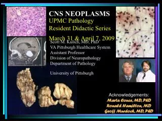
CNS NEOPLASMS. UPMC Pathology Resident Didactic Series March 31 & April 7, 2009. Scott M. Kulich, MD, PhD VA Pittsburgh Healthcare System Assistant Professor Division of Neuropathology Department of Pathology University of Pittsburgh. Acknowledgements: Marta Couce, MD, PhD

E N D
CNS NEOPLASMS UPMC Pathology Resident Didactic SeriesMarch 31 & April 7, 2009 Scott M. Kulich, MD, PhDVA Pittsburgh Healthcare SystemAssistant ProfessorDivision of NeuropathologyDepartment of PathologyUniversity of Pittsburgh Acknowledgements: Marta Couce, MD, PhD Ronald Hamilton, MD Geoff Murdoch, MD, PhD
Outline • Neuroradiology for pathologists • Familial tumor syndromes • CNS neoplasms • Astrocytic neoplasms • Diffuse astrocytomas -> GBM • Variants • Pilocytic astrocytomas • Pleomorphic xanthoastrocytoma • Subependymal giant cell astrocytoma • Oligodendrogliomas • Oligoastrocytomas • Other neuroepithelial • Angiocentric glioma, chordoid glioma, astroblastoma • Ependymomas
Outline (CNS neoplasms cont.) • Choroid plexus • Neuronal - Neuroglial Tumors • Ganglioglioma • Central neurocytoma • Paraganglioma • Embryonal tumors • Meningeal tumors
Outline • Neuroradiology for pathologists • Familial tumor syndromes • CNS neoplasms • Astrocytic neoplasms • Diffuse astrocytomas -> GBM • Variants • Pilocytic astrocytomas • Pleomorphic xanthoastrocytoma • Subependymal giant cell astrocytoma • Oligodendrogliomas • Oligoastrocytomas • Other neuroepithelial • Angiocentric glioma, chordoid glioma, astroblastoma • Ependymomas
NEURORADIOLOGY FOR PATHOLOGISTS Question: Who cares?
Answer: You will when your favorite neurosurgeon hands you a piece of tissue the size of a grain of salt and tells you he needs you to tell him if he can go ahead and stick Gliadel chemotherapeutic wafers in the patient’s brain NEURORADIOLOGY FOR PATHOLOGISTS Question: Who cares?
Answer: You will when your favorite neurosurgeon hands you a piece of tissue the size of a grain of salt and tells you he needs you to tell him if he can go ahead and stick Gliadel chemotherapeutic wafers in the patient’s brain NEURORADIOLOGY FOR PATHOLOGISTS Question: Who cares? Neuroradiology = Gross pathology
NEURORADIOLOGY FOR PATHOLOGISTS Neuroradiology for • Two main imaging techniques • Computerized tomography (CT) • 3D X-rays • White areas = areas that absorb or “attenuate” the passage of x-ray beam (acute hematoma, bone, calcium = hyperdense/ attenuating) • Black areas = areas that do not absorb or “attenuate” the passage of x-ray beam (fat, air, CSF, edema = hypodense/ attenuating)
Neuroradiology for
NEURORADIOLOGY FOR PATHOLOGISTS • Magnetic resonance imaging (MRI) • Not ionizing radiation but magnetic field to excite protons which emit “signal” upon relaxation • Image appearance dependent upon time interval between each excitation and time interval between each collection • Two basic “weights” of images based upon TE and TR • T1: Short TE and TR • T1 is the one…that looks like a brain • T2 :Long TE and TR
NEURORADIOLOGY FOR PATHOLOGISTS • Magnetic resonance imaging (MRI) • Not ionizing radiation but magnetic field to excite protons which emit “signal” upon relaxation • Image appearance dependent upon time interval between each excitation and time interval between each collection • Two basic “weights” of images based upon TE and TR • T1: Short TE and TR • T1 is the one…that looks like a brain • T2 :Long TE and TR
NEURORADIOLOGY FOR PATHOLOGISTS • Magnetic resonance imaging (MRI) • Not ionizing radiation but magnetic field to excite protons which emit “signal” upon relaxation • Image appearance dependent upon time interval between each excitation and time interval between each collection • Two basic “weights” of images based upon TE and TR • T1: Short TE and TR • T1 is the one…that looks like a brain • T2 :Long TE and TR
NEURORADIOLOGY FOR PATHOLOGISTS • Important info to glean from neuroimaging • Age • Location, location, location • Multicentricity • Bilateral hemisphere involvement • Architecture • Contrast enhancement • Interaction with surrounding tissue
Location, location, location… CHILDREN
NEURORADIOLOGY FOR PATHOLOGISTS • Multicentricity • Neoplasms • Metastatic disease • Others (lymphoma, high-grade glioma,…) • Non-neoplastic • Demyelinating disease • Infectious • Bilateral hemisphere involvement • “butterfly” lesion • Glioblastoma multiforme (GBM), lymphoma
NEURORADIOLOGY FOR PATHOLOGISTS • Multicentricity • Neoplasms • Metastatic disease • Others (lymphoma, high-grade glioma,…) • Non-neoplastic • Demyelinating disease • Infectious • Bilateral hemisphere involvement • “butterfly” lesion • Glioblastoma multiforme (GBM), lymphoma
NEURORADIOLOGY FOR PATHOLOGISTS • Architecture • CYSTIC = LOW-GRADE • JPA (juvenile pilocytic astrocytoma), PXA (pleomorphic xanthoastrocytoma), ganglion cell tumors, • Others (hemangioblastoma, craniopharygioma, supratentorial ependymomas, extraventricular neurocytoma) • Frequently associated with a mural nodule (JPA, PXA, hemangioblastoma, ganglion cell tumors,PGNT, extraventricular neurocytoma) • Dural tail • Meningioma
NEURORADIOLOGY FOR PATHOLOGISTS • Architecture • CYSTIC = LOW-GRADE • JPA (juvenile pilocytic astrocytoma), PXA (pleomorphic xanthoastrocytoma), ganglion cell tumors, • Others (hemangioblastoma, craniopharygioma, supratentorial ependymomas, extraventricular neurocytoma) • Frequently associated with a mural nodule (JPA, PXA, hemangioblastoma, ganglion cell tumors,PGNT, extraventricular neurocytoma) • Dural tail • Meningioma
NEURORADIOLOGY FOR PATHOLOGISTS • Contrast enhancement • Breached blood-brain barrier • Seen with neoplasms but can be seen with other conditions (e.g. infectious, demyelinating, …) • Pattern of enhancement often helpful • Homogeneous versus non-homogeneous • Lymphoma, hemangiopericytoma, meningioma • GBM, mets, abscesses • Patchy versus circumferential ( i.e. ring enhancement)
NEURORADIOLOGY FOR PATHOLOGISTS • Contrast enhancement • Breached blood-brain barrier • Seen with neoplasms but can be seen with other conditions (e.g. infectious, demyelinating, …) • Pattern of enhancement often helpful • Homogeneous versus non-homogeneous • Lymphoma, hemangiopericytoma, meningioma • GBM, mets, abscesses • Patchy versus circumferential ( i.e. ring enhancement)
NEURORADIOLOGY FOR PATHOLOGISTS Heterogeneous enhancement (GBM)
NEURORADIOLOGY FOR PATHOLOGISTS Homogeneous enhancement (Meningioma)
NEURORADIOLOGY FOR PATHOLOGISTS • Interaction with surrounding tissue • Edema • “Activity” of lesion • Malignant neoplasms • Inflammatory lesions • Skull • Erosion: Long-standing low-grade lesions • Dysembryoplastic neuroepithelial tumor (DNET), PXA, ganglion cell tumors,oligodendrogliomas,epidermoid cysts • Hyperostosis • Meningiomas
NEURORADIOLOGY FOR PATHOLOGISTS • Interaction with surrounding tissue • Edema • “Activity” of lesion • Malignant neoplasms • Inflammatory lesions • Skull • Erosion: Long-standing low-grade lesions • Dysembryoplastic neuroepithelial tumor (DNET), PXA, ganglion cell tumors,oligodendrogliomas,epidermoid cysts • Hyperostosis • Meningiomas
NEURORADIOLOGY FOR PATHOLOGISTS • Interaction with surrounding tissue • Edema • “Activity” of lesion • Malignant neoplasms • Inflammatory lesions • Skull • Erosion: Long-standing low-grade lesions • Dysembryoplastic neuroepithelial tumor (DNET), PXA, ganglion cell tumors,oligodendrogliomas,epidermoid cysts • Hyperostosis • Meningiomas
Approach to intraoperative consults • Review of imaging and history • Questions for surgeon • What do you NEED to know? • Can you get more tissue if necessary? • Specimen preparation • Intraoperative cytology vs frozen sections • touch and smear preparations
Approach to intraoperative consults • Review of imaging and history • Questions for surgeon • What do you NEED to know? • Can you get more tissue if necessary? • Specimen preparation • Intraoperative cytology vs frozen sections • touch and smear preparations
Approach to intraoperative consults • Review of imaging and history • Questions for surgeon • What do you NEED to know? • Can you get more tissue if necessary? • Specimen preparation • Intraoperative cytology vs frozen sections • touch and smear preparations
Approach to intraoperative consults • Specimen preparation • Intraoperative cytology • Smear preparations
Approach to intraoperative consults • Specimen preparation • Intraoperative cytology • Smear preparations
A “wiley” approach to intraoperative consults • Abnormal versus normal • Reactive versus neoplastic • Primary versus metastatic • Grade of lesion • Does diagnosis correlate with clinical and imaging data?
A “wiley” approach to intraoperative consults • Abnormal versus normal • Reactive versus neoplastic • Primary versus metastatic • Grade of lesion • Does diagnosis correlate with clinical and imaging data?
A “wiley” approach to intraoperative consults • Abnormal versus normal • Reactive versus neoplastic • Primary versus metastatic • Grade of lesion • Does diagnosis correlate with clinical and imaging data?
A “wiley” approach to intraoperative consults • Abnormal versus normal • Reactive versus neoplastic • Primary versus metastatic • Grade of lesion • Does diagnosis correlate with clinical and imaging data?
A “wiley” approach to intraoperative consults • Abnormal versus normal • Reactive versus neoplastic • Primary versus metastatic • Grade of lesion • Does diagnosis correlate with clinical and imaging data?
Kulich Any questions?