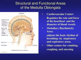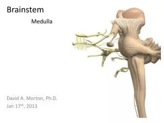
MEDULLA OBLONGATA
250 likes | 485 Vues
Explore the caudal medulla's internal structure, from trigeminal sensory nucleus to dorsal columns and more, in Dr. Vohra's detailed examination.

MEDULLA OBLONGATA
E N D
Presentation Transcript
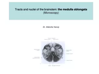
MEDULLA OBLONGATA INTERNAL FEATURES
At the transition from spinal cord to medulla, the pattern of grey and white matter undergoes considerable rearrangement. The ventral horn becomes much attenuated The dorsal horn is replaced by the caudal part of the trigeminal sensory nucleus (nucleus of the spinal tract of the trigeminal nerve). Dr. Vohra
TRIGEMINAL SENSORY NUCLEUS • The trigeminal sensory nucleus is regarded as the brain stem homologue of the dorsal horn since it receives primary afferent fibres conveying general sensation from the head, which enter the brain stem in the trigeminal nerve. • It is a large nucleus that extends the whole length of the brain stem and into the upper segments of the spinal cord. • This latter, caudal part of the trigeminal nucleus is particularly associated with the modalities of pain and temperature. Dr. Vohra
SPINAL TRACT OF THE TRIGEMINAL • The trigeminal nerve attaches to the pons. • Fibres that terminate in the parts of the trigeminal nucleus caudal to this level descend in a tract (the spinal tract of the trigeminal)which lies immediately superficial to the nucleus. Dr. Vohra
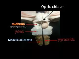
DECUSSATION OF PYRAMIDS In the ventral medulla, the majority of fibres of the pyramid undergo decussation then pass laterally, dorsally and caudally to form the lateral corticospinal tract. Decuss- = crossing Dr. Vohra
VENTRAL SURFACE On the ventral surface of the mid medulla the pyramids are prominent, above their decussation. Dr. Vohra
DORSAL SURFACE On the dorsal surface, the dorsal columns reach their termination in the gracile and cuneate nuclei which appear beneath their respective tracts. • The dorsal columns consist of first-order sensory neurones; the cell bodies of these neurones lie in the dorsal root ganglia of spinal nerves and have central processes that ascended ipsilaterally through the cord and into the medulla. • They terminate in the nucleus gracilis and cuneatus upon the cell bodies of second-order neurones. Dr. Vohra
MEDIAL LEMNISCUS • The axons of these neurones course ventrally and medially as internal arcuatefibres, decussating in the midline. • Thereafter, they turn rostrally forming a distinct tract, the medial lemniscus, that runs through the rostral medulla, the pons and midbrain to terminate in the ventral posterior nucleus of the thalamus. lemniscus = ribbon Dr. Vohra
VENTRAL SURFACE • Dorsolateral to the pyramid and lateral to the medial lemniscus is the inferior olivary nucleus,lying within the prominence of the olive. • On the ventral surface of the medulla, the pyramids remain conspicuous. • Immediately dorsal to the medial aspect of the pyramid lies the medial lemniscus, on either side of the midline. Dr. Vohra
INFERIOR OLIVARY NUCLEUS • The inferior olivary nucleus has the appearance of a crenated bag with an opening, or hilum, facing medially and through which afferent and efferent fibres pass. • It is concerned with the control of movement and receives afferents from the motor and sensory cortices of the cerebral hemisphere and from the red nucleus of the midbrain. Dr. Vohra
INFERIOR OLIVARY NUCLEUS • Its main efferent connection is to the cerebellum via the inferior cerebellar peduncle. • Within the cerebellum its axons, known as climbing fibres, end in excitatory synapses in the dentate nucleus and upon Purkinje cells of the cerebellar cortex. Dr. Vohra
Dorsal to the inferior olivary nucleus and lateral to the medial lemniscus lie second-order sensory fibres ascending to the ventral posterior thalamus. They come from the trigeminal nucleus (the trigeminothalamic tract) and from the spinal cord (spinothalamic fibres, referred to in the brain stem as the spinal lemniscus). Dr. Vohra
HYPOGLOSSAL NUCLEUS • The dorsal surface of the rostral medulla forms part of the floor of the fourth ventricle. Both immediately and deep beneath the floor of the ventricle lie a number of cranial nerve nuclei, some of which can be clearly identified in simply stained sections, others of whichcannot. • Immediately beneath the ventricular floor, just lateral to the midline, lies the hypoglossal nucleus, which contains motor neurones innervating the muscles of the tongue via the hypoglossal nerve. Dr. Vohra
VAGAL NUCLEUS • Lateral to the hypoglossal nucleus lies the dorsal (motor) nucleus of the vagus, containing preganglionic parasympathetic neurones that run in the vagus nerve. • The most caudal aspect of the ventricular floor is known as the area postrema. • At this point the blood-brain barrier which limits the passage of certain chemicals from the blood to the brain, is absent. • This region is the central site of action of substances that cause vomiting (emetics). • In the lateral part of the floor of the fourth ventricle are located the vestibular nuclei,which receive primary afferent fibres from the vestibular nerve. Dr. Vohra
MEDIAL LONGITUDINAL FASCICULUS • Ventromedial to the hypoglossal nucleus, close to the midline, is located the medial longitudinal fasciculus. • This consists of both ascending and descendingfibres and can be identified also in the pons and mid-brain. • Within the brain stem, it links the vestibular nuclei with the nuclei supplying the extraocular muscles (abducens, trochlear and oculomotor nuclei) and subserves the coordination of head and eye movements. Dr. Vohra
RESTIFORM BODY • The dorsolateral part of the rostral medulla is dominated by the inferior cerebellar peduncle, or restiform body.This consists of fibres passing between the medulla and the cerebellum. • Prominent amongst these are olivocerebellarfibres, connections between the vestibular nuclei and the cerebellum, and the fibres of the dorsal spinocerebellar tract, conveying proprioceptive information from the limbs. Dr. Vohra
COCHLEAR AND AMBIGUUS NUCLEI • On the dorsal and lateral aspects of the inferior cerebellar peduncle lie the dorsal and ventral cochlear nuclei, which receive afferent information from the cochlear nerve. • Deep beneath the ventricular floor, just dorsal to the inferior olivary nucleus, is located the nucleus ambiguus. • This sends motor fibers into the glossopharyngeal, vagus and accessory nerves and, thence, to the muscles of the pharynx and larynx. Dr. Vohra