Cell Cycle & Division Mechanism
310 likes | 354 Vues
Explore cell growth, chromosomal cycles, mitosis stages, and the process of meiosis. Learn about the phases involved in cell division and the significance of each stage in cellular reproduction.

Cell Cycle & Division Mechanism
E N D
Presentation Transcript
Cell cycle and cell division (THE MECHANISM OF CELL DIVISION)
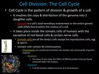
INTRODUCTION: • Two types of organism- Unicellular & multicellular • Multicellular organisms are made up of two types of cells i.e. somatic/body cells & germinal cells • somatic cells form the body of organism while the germ cells form sex cells/ gametes • Growth and development depends on growth and multiplication of the cells
Growth patterns are of 3 types of- • 1. Auxetic growth: An increase of cell mass • 2. Multiplicative growth: An increase in cell number by multiplication • 3. Accretionary growth: Due to accumulation of extracellular products.
History: • Virchow (1855) who properly studied the cell division • Animal cell division studied by Prevost and Dumas in 1824 • Remak Kolliker(1841) showed that the processes involves a division of both nucleus and cytoplasm • The term karyokinesis was introduced by Schleicher1878, • while cytokinesis by Whitermanin 1887
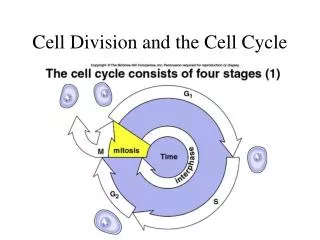
CELL CYCLE • A dividing cell shows number of physical and chemical changes (metabolic changes) before, during and after cell division. • All such changes together constitute cell cycle • These changes are orderly & sequence is common for all the cells in all organisms • Cell cycle can be defined as “the entire sequence of events happening from end of one nuclear division to the beginning of the next”
Cell cycle involves following 3 cycles • Chromosomal cycle: DNA synthesis alternates with mitosis. During DNA synthesis each double helical DNA is replicated in to two identical daughter DNA. And during mitosis the duplicated copies of genome are separated • Cytoplasmic cycle: Usually karyokinesis is followed by cytokinesis to form the two poles of the mitotic spindle 3. Centrosome cycle: both the above cycles require that the centrosome be inherited and duplicated in order.
Howard & Pelc(1953) divided cell cycle in to 4 phases, viz. G1phase, S phase ,G2 phase and M Phase.
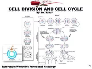
MitosisIt is a process of cell division which results in the production of two daughter cells from a single parent cell. The daughter cells are identical to one another and to the original parent cell.In a typical animal cell, mitosis can be divided into four principals stages:
Definition: Mitosis is defined as the type of cell division by which a single cell divides in such a way as to produce two genetically identical "daughter cells". This is the method by which the body produces new cells for both growth and repair of aging or damaged tissues throughout the body - as opposed to for sexual reproduction Mitosis is the simplest of the two ways (mitosis and meiosis) in which the nucleus of a cell can divide - as part of a process of whole cell division. The four stages of mitosis (prophase, metaphase, anaphase and telophase) are illustrated and described below.
ProphaseEarly in the prophase stage the chromatin fibres shorten into chromosomes that are visible under a light microscope. (Each prophase chromosome consists of a pair of identical double-stranded chromatids.) Later in prophase, the nucleolus disappears, the nuclear envelope breaks down, and the two centrosomes begin to form the miotic spindle (which is an assembly of microtubules). As the microtubules extend in length between the centrosomes, the centrosomes are pushed to opposite "poles" (extremes) of the cell. Eventually, the spindle extends between two opposite poles of the cell.
1) Prophase: The chromatin, diffuse in interphase, condenses into chromosomes. Each chromosome has duplicated and now consists of two sister chromatids. At the end of prophase, the nuclear envelope breaks down into vesicles.2) Metaphase: The chromosomes align at the equatorial plate and are held in place by microtubules attached to the mitotic spindle and to part of the centromere. 3) Anaphase: The centromeres divide. Sister chromatids separate and move toward the corresponding poles.
4. Telophase: Daughter chromosomes arrive at the poles and the microtubules disappear. The condensed chromatin expands and the nuclear envelope reappears. The cytoplasm divides, the cell membrane pinches inward ultimately producing two daughter cells (phase: Cytokinesis).
MEIOSIS:Illustration of the process by which a single parent diploid cell (Both homologous chromosomes) divides to produce four daughter haploids cells (One homologous chromosome of the pair).Meiosis is thetype of cell division by which germ cells (eggs and sperm) are produced. Meiosis involves a reduction in the amount of genetic material.Meiosis comprises two successive nuclear divisions with only one round of DNA replication. Four stages can be described for each nuclear division.
Interphase:Before meiosis begins, genetic material is duplicated. A) First division of meiosis Prophase 1: Duplicated chromatin condenses. Each chromosome consists of two, closely associated sister chromatids. Crossing-over can occur during the latter part of this stage. (stages- L.Z,P,D,D)Metaphase 1: Homologous chromosomes align at the equatorial plate. Anaphase 1: Homologous pairs separate with sister chromatids remaining together. Telophase 1: Two daughter cells are formed with each daughter containing only one chromosome of the homologous pair.
B) Second division of meiosis: Gamete formationProphase 2: DNA does not replicate. Metaphase 2: Chromosomes align at the equatorial plate. Anaphase 2: Centromeres divide and sister chromatids migrate separately to each pole. Telophase 2: Cell division is complete. Four haploid daughter cells are obtained.
One parent cell produces four daughter cells.Daughter cells have half the number of chromosomes found in the original parent cell and with crossing over, are genetically different.Meiosis differs from mitosis primarily because there are two cell divisions in meiosis, resulting in cells with a haploid number of chromosomes.