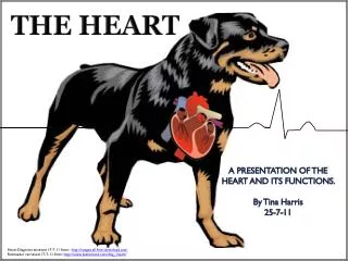
The Heart
The Heart. At the very heart of things is your heart. The heart is the pump of the circulatory system Functions of circulatory system Blood carries oxygen and nutrients to cells Carries waste products away. Organs of the circulatory system. Heart- Arteries Thick walled Veins Thin walled


The Heart
E N D
Presentation Transcript
The Heart At the very heart of things is your heart
The heart is the pump of the circulatory system • Functions of circulatory system • Blood carries oxygen and nutrients to cells • Carries waste products away
Organs of the circulatory system • Heart- • Arteries • Thick walled • Veins • Thin walled • Capillaries • Walls are one cell thick to permit the exchange of gas, waste
Arteries carry blood away from the heart • Veins carry blood back to the heart • There are two major blood circuits • General (systemic) circulation: throughout the body • Cardiopulmonary: from heart to lungs, and back
Location: Thoracic cavity, middle of chest. Between the lungs, above the diaphragm • About the size of a closed fist • 5 inches long, 3.5 wide, weighing less than a pound
Structure • Encased in the pericardium- double layer of fibrous tissue. Between the layers is a lubricating fluid called pericardial fluid • Myocardium- cardiac muscle tissue • Septum- a thick muscular wall separating the right half from the left
Two upper chambers are the atria- right atrium and left atrium • Two lower chambers are the ventricles- right and left. • The deoxygenated blood enters the right side of the heart through the superior and inferior vena cava
Tricuspid valve located between the right atria and ventricle. Cordae tendinae keep it closed. Blood flows from the right ventricle through the pulmonary artery. The pulmonary semi-lunar valve keeps blood from flowing back into the ventricle Pulmonary artery takes deoxygenated blood to the lungs
The return trip • Oxygenated blood is carried back from the lungs via the pulmonary veins- right and left. • Both deposit oxygenated blood into the left atria. When this blood flows into the left ventricle, it is the bicuspid valve that snaps shut between the left atria and ventricle
Oxygenated blood leaves the left side of the heart through the massive contraction of the left ventricle, via the aorta. The aortic semi-lunar valve keeps blood flowing in the right direction • Aorta is the body’s largest vessel-about the size of a garden hose!
The heart is a side-by-side double pump with 4 chambers. Each time the right ventricle contacts to send blood to the lungs pulmonary artery, the right ventricle contracts sending blood into the aorta. • The familiar lubb-dupp are the valves closing
Control of heart contractions • A heart removed from the body will continue to beat- the heart has tissue which controls its own beating! • This group of tissue is where the superior vena cava joins the right atrium- called the sinoatrial node or pacemaker
The SA node sends out an electrical signal- atria contract or depolarize. • Signal travels from the SA node to the AV node, down through the AV bundle. AV bundle further divides into smaller units-Perkinje fibers. Ventricles contract.
Contraction = depolarization= systole • Relaxation = repolarization= diastole • The cardiac cycle takes 0.8 seconds- the average heart beats 72-80 times per minute
The electrical activity can be tracked with an electrocardiogram, a.k.a. EKG or ECG • P wave = atrial depolarization • QRS = ventricular depolarization • T wave = ventricular repolarization
Diagnostic tests • Cardiac catheterization- uses a catheter to deliver dye to heart’s vascular system • Stress test- EKG on a treadmill • Echocardiogram • Holter monitor • Troponin, CPK and LDH enzymes
Symptoms of heart disease • Arrhythmia- irregular heart beat • Bradycardia- slow heart beat <60 beats/min • Tachycardia- rapid heart beat >100 beats/min • Murmur-defect in the valves of the heart • Systolic murmur • Diastolic murmur
Heart disease • Myocardial infarction, a.k.a an MI or heart attack, is caused by a lack of blood to the heart muscle -the myocardium • Causes: arteriosclerosis (hardening of the arteries) • Arteriolosclerosis: collagen • Atherosclerosis: cholesterol plaque
Result; damage to the heart muscle due to lack of oxygen • Symptoms: crushing, severe chest pain radiating to the left shoulder, arm, neck and jaw. Nausea, increased perspiration, fatigue and dyspnea. • Treatment: clot dissolving drugs such as tPA (tissue plasminogen activator), anticoagulants and digitalis • Angioplasty and bypass surgery may be considered