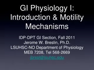
GI Physiology I: Introduction & Motility Mechanisms
560 likes | 1.01k Vues
GI Physiology I: Introduction & Motility Mechanisms. IDP-DPT GI Section, Fall 2011 Jerome W. Breslin, Ph.D. LSUHSC-NO Department of Physiology MEB 7208, Tel 568-2669 jbresl@lsuhsc.edu. Lecture 1 Outline. Introduction to GI Physiology Overview of the Functional Anatomy of the GI Tract.

GI Physiology I: Introduction & Motility Mechanisms
E N D
Presentation Transcript
GI Physiology I:Introduction & Motility Mechanisms • IDP-DPT GI Section, Fall 2011 • Jerome W. Breslin, Ph.D. • LSUHSC-NO Department of Physiology • MEB 7208, Tel 568-2669 • jbresl@lsuhsc.edu
Lecture 1 Outline • Introduction to GI Physiology • Overview of the Functional Anatomy of the GI Tract. • Functions of the GI System. • Processes in the GI Tract. • Water and Solids Balance. • Enteric Nervous System • Immune Function in GI System • Splanchnic Circulation • Motility • Motility Patterns • Basic Mechanisms Underlying Motility
Required Reading: • Gastrointestinal Physiology, Kim E. Barrett, Chapter 1, Chapter 7 - section on peristalsis, Chapter 8 - sections on innervation, basal electrical rhythm, and motility during fasting. • Suggested Reading: • Review of Medical Physiology, William Ganong, Chapters 26 and 27. • Both are freely available for students online through the LSUHSC library website or www.accessmedicine.com
Single Cell Organisms Diffusion of water and ions, Phagocytosis/Endocytosis of larger particles, digestion & absorption in lysosomes
Multicellular Organisms Shape is important! If the shape is not hollow: Greater Ratio of Volume to Exterior Surface Area than in a Single Cell
Hydra (Image from Wikipedia) Simple Multicellular Organisms Shape is important! Cavity or Lumen for optimal digestion and absorption Organization into shapes that maximize surface area for exchange
More complicated multicellular organisms: Humans 1. Terrestrial - not living in an aqueous solution filled with nutrients. 2. Specialized tube through the body for getting nutrients to the circulatory system for delivery to tissues.
GI Function • Take relatively large, solids or gels, and digest them into smaller molecules that can be absorbed as nutrients, while still serving as a barrier to toxins, bacteria, parasites, etc. • Our overall objective for these lectures is to understand biological mechanisms that facilitate GI function.
GI: Functional Anatomy • GI system is a hollow organ, a tube through the body. • The lumen is “outside” the body’s tissues, but its environment is tightly controlled by the body. • Specialized organs for secretion of enzymes & bile. • Epithelial cells line the entire GI tract and serve as the primary barrier. Specialized epithelial cells also secrete and absorb various compounds to/from the lumen. • Epithelium, mucosa, two layers of smooth muscle, blood vessels and lymphatics, nerves. • Structure maximizes surface area for secretion and absorption (folds, villi, and crypts). • Sphincters regulate movement between segments.
Figure 15-3 Many functions in the gut are found in specific locations along its length. Most of the absorption of nutrients occurs in the small intestine, so most of digestion is accomplished there or upstream.
General Anatomy of Gut Wall Figure 15-6 (Contains connective tissue, immune cells, capillaries, nerve endings) (Might have role in villus movement) The gut wall has a layered organization, with the absorptive cells lining the lumen and neural and muscular components below. Blood and lymph vasculature is abundant to transport absorbed nutrients. See Fig. 1-2 and Fig. 1-3 in Barrett’s book,
Folds in the small intestine increase surface area for exchange: Fold of Kerckring Fig. 14-56 from Wilson et al, Histology Image Review Folds of Kerckring, a.k.a. valvulae conniventes
Villi & Crypts Vander, Fig. 15-7 Ganong, Fig. 26-27
Microvilli on luminal surface of intestinal epithelial cells
Degree by which different anatomical features increase surface area in the small intestine: Increase in Surface Area (Relative to cylinder) Structure Surface Area (cm2) Area of simple cylinder 1 ~3,300 4 cm Dia. x 260 cm L Folds of Kercking 3 ~10,000 Villi 30 ~100,000 Microvilli 600 ~2,000,000
GI Sphincters Unitary smooth muscle rings that act as valves Also see Fig. 1-4 in Barrett
GI Sphincters Unitary smooth muscle rings that act as valves • Resting State • Pressure in sphincter > adjacent segments • Inhibits movement between segments • Relaxation • Pressure in sphincter = adjacent segments • Allows forward flow • Constriction • Pressure in sphincter >> adjacent segments • Prevents retrograde flow
Figure 15-5 Water and Solids Balance Digestive secretions are mostly water, with the average amounts indicated here. Note that only 100 ml are excreted in feces, so the mechanisms for water absorption are efficient (recall the kidneys’ primary role in water and osmotic homeostasis).
Innervation of the GI System • Autonomic NS • Parasympathetic Fibers • Sympathetic Fibers • Enteric Nervous System
Enteric Nervous System • Intrinsic Control of the GI Tract: GI Reflexes • Can act independently of the CNS. Local Reflexes = “Short Reflexes.” • Cholinergic and Adrenergic Neurons. • Can be influenced by CNS. “Long Reflexes.”
Figure 15-13 Enteric Nervous System The enteric nervous system coordinates digestion, secretion, and motility to optimize nutrient absorption. Its activity is modified by information from the CNS and from local chemical and mechanical sensors.
Structure of Enteric Nervous System in Gut Wall Fig. 1-8 in Barrett.
Immune Function in the GI System • Gut Associated Lymphoid Tissue (GALT), including Peyer’s Patches in the lamina propria of small intestine. • Immune surveillance for potential pathogens in the small intestine. • Contains macrophages, dendritic cells, B lymphocytes, and T lymphocytes. • M Cells in the epithelium - antigen presenting cells that encounter and present antigens to B and T lymphocytes.
Gastrointestinal System: Processes • Motility • Digestion • Secretion • Absorption Ingestion, Swallowing, Peristalsis, Elimination Physical (Chewing & Grinding) Chemical (Digestive Enzymes) Water, HCl, Enzymes, Some Organic Waste Products Water, Electrolytes, Simple Sugars, Amino Acids, Fatty Acids, Vitamins, Minerals
Motility • Peristalsis = forward propulsion. • Segmental contractions: mixing. • Mouth and Esophagus: Chewing, Swallowing, Peristalsis • Stomach: Filling, Churning, Peristalsis, Emptying • Small Intestine: Segmental Contractions, Peristalsis • Large Intestine: Haustral Shuttling, Mass Movements, Defecation. • Sphincters: Regulation of Movement
GI Smooth Muscle: Circular Muscle and Longitudinal Muscle Berne & Levy, Fig. 31- 4A • Longitudinal Muscle • Thin Muscle Coat • Contraction shortens intestine length & expands radius • Innervated by excitatory motor neurons • Activated by excitatory motor neurons • Few gap junctions to adjacent fibers • Extracellular Ca2+ influx important in excitation-contraction coupling • Circular Muscle • Thick Muscle Coat • Contraction increases intestine length & decreases radius • Innervated by excitatory & inhibitory motor neurons • Activated by myogenic pacemakers & excitatory motor neurons • Many gap junctions to adjacent fibers • Intracellular Ca2+ release important in excitation-contraction coupling
Coordinated, directional contraction of smooth muscle propels ingested food forward (Peristalsis) See Fig. 7-3 in Barrett.
Peristalsis • Propulsive contraction of the circular muscle • Evoked by distention of intestinal wall • Does not occur after paralysis of ENS • Longitudinal muscle ahead of bolus contracts, circular muscle layer relaxes, and segment receives the aborally moving intestinal contents • Circular muscle behind bolus contracts, longitudinal muscle simultaneously relaxes • Provides propulsive force necessary to move the contents into the receiving segment
Figure 15-32 Segmental Contractions: Mixing Most of the contractions of the small intestine are of the mixing and churning actions portrayed here as segmentation contractions; peristalsis and the downstream movement of materials is infrequent.
Mixing Movements • Mix chyme with digestive enzymes and increase contact between intraluminal contents and the epithelium for final digestion and absorption • Non-propagating Circular muscle contractions • Circular muscles on either side of contracting band remain relaxed, i.e., receiving segments on both sides of the zone of contraction, resulting in propagation of intestinal contents in both an oral and aboral direction
Myogenic Basis of GI phasic contraction: Slow Waves/Basal Electrical Rhythm Phase: 0 – Resting membrane potential Outward K+ current 1 – Upstroke Depolarization Activation of voltage-dependent Ca2+ channels 2 – Transient Repolarization Inactivation of voltage-dependent Ca2+ channels Activation of voltage gated K+ channels 3 – Plateau Phase Balance of inward Ca2+ current and outward K+ currents 4 – Repolarization Inactivation of voltage-dependent Ca2+ channels Activation of Ca2+-gated K+ channels
Rhythmic waves of smooth muscle contraction in the gut are the result of waves of action potentials moving along via gap junctions. Fig. 8-3 in Barrett
Origin of Phasic and Tonic Contractions Modulate Contractions Origin and Control of GI Motor Function • Myogenic • Smooth Muscle • Interstitial Cells of Cajal • Neurogenic • Intrinsic (Enteric NS) • Extrinsic (SNS and PNS) • Endocrine • Paracrine
Slow Waves = Basal Electrical Rhythm (BER) • Depolarizations of smooth muscle cells • Controlled by Intersitial Cells of Cajal (ICC) • BER is propogated via gap junctions to a limited number of adjacent cells. • BER is propogated in the aboral direction.
Slow Waves/Basal Electrical Rhythm (BER) Berne & Levy, Fig. 31-6 1. Slow waves only produce contraction when the threshold is achieved. 2. Slow waves determine maximal rhythm of phasic contractions.
Ganong, Fig. 26-2 3. Neurogenic and Endocrine inputs do not alter the BER, but can facilitate reaching the threshold for contraction.
BER in different segments: • Stomach ~ 3/min • Duodenum ~ 11-12/min • Distal Ileum and Colon ~ 6-7/min Note - 3 slides ahead in handout
Interstitial Cells of Cajal (ICC) generate GI slow waves (basal electrical rhythm) Wild-type mouse ICC-deficient mouse ICC-deficient mouse ICC-deficient mouse from Horowitz et al, Annual Review of Physiology, 61:19-43, 1999)
INTERSTITIAL CELLS OF CAJAL (ICC) • Cells mediate between efferent neurons and smooth muscle cells: • Responsible for slow waves and pacemaker activity of smooth muscle. • Also amplify neuronal input. • Central to GI motility regulation. • Loss of ICC implicated in many human motility disorders (Hirshsprung’s disease, severe constipation, IBD, etc). • Current evidence suggests that mechanism involves Ca++ release from IP3-operated stores – this triggers Ca++ uptake by mitochondrialeading to generation of pacemaker currents.
GI Motility: Neural and Endocrine Inputs • BER is minimally affected by neural and endocrine influences. It is intrinsic to ICC and SM. • However, neurogenic and endocrine stimuli can influence membrane potential of SM, • For example, ACh will enhance depolarization, formation of spike potentials, and SM contraction.