Diffusion Physics
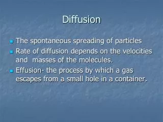
Diffusion Physics. H 2 O in the body is always in random motion due to thermal agitation. B.M. Ellingson, Ph.D., Dept. of Radiological Sciences, David Geffen School of Medicine at UCLA, 2010. Diffusion Physics. Diffusion Coefficient is dependent on Temperature and Viscosity of Tissue.

Diffusion Physics
E N D
Presentation Transcript
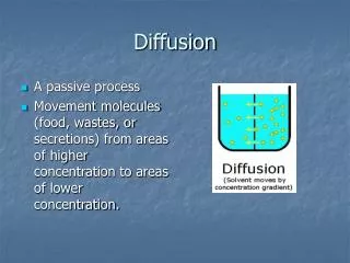
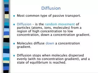
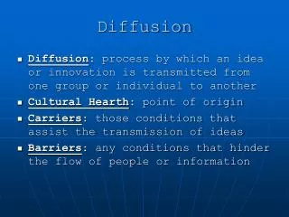
Diffusion Physics • H2O in the body is always in random motion due to thermal agitation B.M. Ellingson, Ph.D., Dept. of Radiological Sciences, David Geffen School of Medicine at UCLA, 2010
Diffusion Physics • Diffusion Coefficient is dependent on Temperature and Viscosity of Tissue Diffusion Coefficient (rate of motion) Temperature Viscosity Size of Molecule B.M. Ellingson, Ph.D., Dept. of Radiological Sciences, David Geffen School of Medicine at UCLA, 2010
Diffusion Physics • The “rate” of water motion is determine by a diffusion coefficient, “D”. • Mean displacement of water molecules is related to “D” by Einstein’s equation: Mean Displacement Diffusion Coefficient Time B.M. Ellingson, Ph.D., Dept. of Radiological Sciences, David Geffen School of Medicine at UCLA, 2010
Detecting Diffusion with MRI - Intravoxel Incoherent Motion Ellingson et al., Concepts in MR, 2008 From: Ellingson, Concepts in MR, 2008 B.M. Ellingson, Ph.D., Dept. of Radiological Sciences, David Geffen School of Medicine at UCLA, 2010
Detecting Diffusion with MRI - Intravoxel Incoherent Motion Variability in Phase of “Tagged” H2O Level of Diffusion Weighting Detected DWI Signal Diffusion Coefficient MRI Signal w/o Diffusion Sensitivity B.M. Ellingson, Ph.D., Dept. of Radiological Sciences, David Geffen School of Medicine at UCLA, 2010
Intravoxel Incoherent vs Coherent Motion Diffusion Effects (Incoherent) Flow Effects (Coherent) -- Phase Contrast (PC)-MRI d = /3 radians Velocity B.M. Ellingson, Ph.D., Dept. of Radiological Sciences, David Geffen School of Medicine at UCLA, 2010
Image Voxel Proton on H2O = t3 = t2 = t1 MRI Signal Diffusion Time (or level of diffusion weighting) B.M. Ellingson, Ph.D., Dept. of Radiological Sciences, David Geffen School of Medicine at UCLA, 2010
Factors that affect diffusion coefficient, D • Diffusion Time, t • Physical time between gradients used to “tag” H2O B.M. Ellingson, Ph.D., Dept. of Radiological Sciences, David Geffen School of Medicine at UCLA, 2010
Factors that affect diffusion coefficient, D • Size of Compartment(s) • If we set a limit for r, then we observe an apparent diffusion coefficient, ADC Physical Compartment Size Expected Compartment Size B.M. Ellingson, Ph.D., Dept. of Radiological Sciences, David Geffen School of Medicine at UCLA, 2010
Factors that affect diffusion coefficient, D • Tortuosity of the Compartments • - More tortuous paths look like slow diffusion Actual Path Tortuosity Expected Path B.M. Ellingson, Ph.D., Dept. of Radiological Sciences, David Geffen School of Medicine at UCLA, 2010
Factors that affect diffusion coefficient, D • Viscosity and Temperature Diffusion Coefficient (rate of motion) Temperature Pure H2O CSF Infection (WBCs) Lymphoma Viscosity B.M. Ellingson, Ph.D., Dept. of Radiological Sciences, David Geffen School of Medicine at UCLA, 2010
Factors that affect diffusion coefficient, D • Diffusion Time, t • Physical time between gradients used to “tag” H2O • Size of Compartment(s) • If we set a limit for r, then we observe an apparent diffusion coefficient, ADC • Tortuosity of the Compartments • - More tortuous paths look like slow diffusion • Temperature • Viscosity *** We can only measure “ADC” because of all the factors that change “D”! *** B.M. Ellingson, Ph.D., Dept. of Radiological Sciences, David Geffen School of Medicine at UCLA, 2010
Steps in Performing DWI • DWI (isotropic): • Collect a DWI (b = 1000 or 500 s/mm2) dataset by applying motion probing gradients in the x, y, and z-direction. • Make sure TE is low and TR is long to increase SNR • For higher resolution scans, use a lower b-value • Collect a T2w dataset (b = 0 s/mm2) • Collect a low b-value, flow nulled dataset (b = 50 s/mm2) • Average DWIs from 3 directions • Calculate ADC For b-values < 1000 B.M. Ellingson, Ph.D., Dept. of Radiological Sciences, David Geffen School of Medicine at UCLA, 2010
DWI vs. ADC • DWI • Images collected during application of a “diffusion sensitizing gradient” • Contains T1, T2, and ADC effects • “Restricted diffusion”, long T2, and short T1 all influence DWIs b = 750 b = 1000 From: Taouli, Radiology, 2010 B.M. Ellingson, Ph.D., Dept. of Radiological Sciences, David Geffen School of Medicine at UCLA, 2010
DWI vs. ADC ADC Map b = 500 • DWI • Influence of T2 in DWIs is known as “T2 shine through” From: Taouli, Radiology, 2010 B.M. Ellingson, Ph.D., Dept. of Radiological Sciences, David Geffen School of Medicine at UCLA, 2010
DWI vs. ADC • ADC • Quantitative • Calculated from DWI and T2w (b = 0 or low b-value) From: Taouli, Radiology, 2010 B.M. Ellingson, Ph.D., Dept. of Radiological Sciences, David Geffen School of Medicine at UCLA, 2010
DWI vs. ADC • ADC • Reflects diffusion magnitude • Eliminates long T2 and short T1 effects From: Taouli, Radiology, 2010 B.M. Ellingson, Ph.D., Dept. of Radiological Sciences, David Geffen School of Medicine at UCLA, 2010
DWI vs. ADC ADC Map b = 500 • DWI • Influence of T2 in DWIs is known as “T2 shine through” From: Taouli, Radiology, 2010 B.M. Ellingson, Ph.D., Dept. of Radiological Sciences, David Geffen School of Medicine at UCLA, 2010
Diffusion Tensor Imaging (DTI) Isotropic Diffusion Anisotropic Diffusion From: Ellingson, Concepts in MR, 2008 B.M. Ellingson, Ph.D., Dept. of Radiological Sciences, David Geffen School of Medicine at UCLA, 2010
Diffusion Tensor Imaging (DTI) Directional Encoding 6 directions (min) 15 directions 25 directions 41 directions B.M. Ellingson, Ph.D., Dept. of Radiological Sciences, David Geffen School of Medicine at UCLA, 2010
Diffusion Tensor Imaging (DTI) The Diffusion Tensor: B.M. Ellingson, Ph.D., Dept. of Radiological Sciences, David Geffen School of Medicine at UCLA, 2010
Diffusion Tensor Imaging (DTI) From Ellingson, Concepts in MR, 2008 Isotropic Diffusion 1 = 2 = 3 Anisotropic Diffusion 1 > 2,3 B.M. Ellingson, Ph.D., Dept. of Radiological Sciences, David Geffen School of Medicine at UCLA, 2010
FA = 0 FA = 1 Isotropic Anisotropic Fractional Anisotropy (FA) B.M. Ellingson, Ph.D., Dept. of Radiological Sciences, David Geffen School of Medicine at UCLA, 2010
DTI Tractography • In the CNS and MSK, lADC is parallel to axon orientation B.M. Ellingson, Ph.D., Dept. of Radiological Sciences, David Geffen School of Medicine at UCLA, 2010
DTI Tractography • In the CNS, lADC is parallel to axon orientation B.M. Ellingson, Ph.D., Dept. of Radiological Sciences, David Geffen School of Medicine at UCLA, 2010
DTI Tractography • In the MSK, lADC is parallel to muscle fiber orientation From: University of Rochester B.M. Ellingson, Ph.D., Dept. of Radiological Sciences, David Geffen School of Medicine at UCLA, 2010
DTI Tractography • In the heart, lADC is also parallel to muscle fiber orientation From: Eindhoven University of Technology From: University of Oxford B.M. Ellingson, Ph.D., Dept. of Radiological Sciences, David Geffen School of Medicine at UCLA, 2010
DTI Tractography • Spinal Cord Injury From Ellingson, Neurosurgery:Spine, 2010 B.M. Ellingson, Ph.D., Dept. of Radiological Sciences, David Geffen School of Medicine at UCLA, 2010
Steps in Performing DTI • DTI (6 directions): • Collect a DWI (b = 1000 or 500 s/mm2) along 6 non-collinear directions • Collect a T2w dataset (b = 0 s/mm2) • Calculate Diffusion Tensor: • Calculate D in 6 different directions • Set up the encoding matrix • Define Tensors • Solve Tensor Equations B.M. Ellingson, Ph.D., Dept. of Radiological Sciences, David Geffen School of Medicine at UCLA, 2010
Applications of Diffusion MRIin the Abdomen B.M. Ellingson, Ph.D., Dept. of Radiological Sciences, David Geffen School of Medicine at UCLA, 2010
Diffusion MR Imagingof the Liver B.M. Ellingson, Ph.D., Dept. of Radiological Sciences, David Geffen School of Medicine at UCLA, 2010
Diffusion MR Imagingof the Liver • Common pulse sequences • Single-shot spin-echo (SE) echoplanar with fat saturation • With breathhold (20-30 seconds; sensitivity for lesion detection = 84.3%Parikh, 2008) • Need thicker slices (8-10 mm) for good SNR and good liver coverage • Resp. gated (3-6 min; sensitivity for lesion detection = 93.7%Parikh, 2008) • Thinner slices can be used (5 mm) • Better image quality, SNR and ADC quantificationTaouli, 2009 B.M. Ellingson, Ph.D., Dept. of Radiological Sciences, David Geffen School of Medicine at UCLA, 2010
Diffusion MR Imagingof the Liver • Common pulse sequences • Single-shot spin-echo (SE) echoplanar with fat saturation • With breathhold (20-30 seconds; sensitivity for lesion detection = 84.3%Parikh, 2008) • Need thicker slices (8-10 mm) for good SNR and good liver coverage • Resp. gated (3-6 min; sensitivity for lesion detection = 93.7%Parikh, 2008) • Thinner slices can be used (5 mm) • Better image quality, SNR and ADC quantificationTaouli, 2009 • Common b-values • b = 0 image (no diffusion weighting…essentially a “poor man’s” T2w image) B.M. Ellingson, Ph.D., Dept. of Radiological Sciences, David Geffen School of Medicine at UCLA, 2010
Diffusion MR Imagingof the Liver • Common pulse sequences • Single-shot spin-echo (SE) echoplanar with fat saturation • With breathhold (20-30 seconds; sensitivity for lesion detection = 84.3%Parikh, 2008) • Need thicker slices (8-10 mm) for good SNR and good liver coverage • Resp. gated (3-6 min; sensitivity for lesion detection = 93.7%Parikh, 2008) • Thinner slices can be used (5 mm) • Better image quality, SNR and ADC quantificationTaouli, 2009 • Common b-values • b = 0 image (baseline with no diffusion weighting…essentially a “poor man’s” T2w image • Low b-value (b < 150 s/mm2) “Flow Nulled” • Nulls the intrahepatic vascular signal • Allows for better detection of focal liver lesions(van den Bos, 2008; Parikh, 2008; Okada, 1998; Hussain, 2005) ~2% change in ADC B.M. Ellingson, Ph.D., Dept. of Radiological Sciences, David Geffen School of Medicine at UCLA, 2010
Diffusion MR Imagingof the Liver • Common pulse sequences • Single-shot spin-echo (SE) echoplanar with fat saturation • With breathhold (20-30 seconds; sensitivity for lesion detection = 84.3%Parikh, 2008) • Need thicker slices (8-10 mm) for good SNR and good liver coverage • Resp. gated (3-6 min; sensitivity for lesion detection = 93.7%Parikh, 2008) • Thinner slices can be used (5 mm) • Better image quality, SNR and ADC quantificationTaouli, 2009 • Common b-values • b = 0 image (baseline with no diffusion weighting…essentially a “poor man’s” T2w image • Low b-value (b < 150 s/mm2) “Flow Nulled” • Nulls the intrahepatic vascular signal • Allows for better detection of focal liver lesions(van den Bos, 2008; Parikh, 2008; Okada, 1998; Hussain, 2005) • High b-value (500 < b < 1000 s/mm2) • Useful for focal liver lesion characterization (Taouli, 2003; Kim, 1999) B.M. Ellingson, Ph.D., Dept. of Radiological Sciences, David Geffen School of Medicine at UCLA, 2010
Diffusion MR Imagingof the Liver • Visual liver lesion characterization with DWI From: Taouli, Radiology, 2010 B.M. Ellingson, Ph.D., Dept. of Radiological Sciences, David Geffen School of Medicine at UCLA, 2010
Diffusion MR Imagingof the Liver • Visual liver lesion characterization with DWI From: Taouli, Radiology, 2010 b = 500 ADC From: Xu, J Comput Assist Tomogr, 2010 B.M. Ellingson, Ph.D., Dept. of Radiological Sciences, David Geffen School of Medicine at UCLA, 2010
Diffusion MR Imagingof the Liver • Visual liver lesion characterization with DWI - DWI has higher specificity than CE From: Xu, J Comput Assist Tomogr, 2010 B.M. Ellingson, Ph.D., Dept. of Radiological Sciences, David Geffen School of Medicine at UCLA, 2010
Diffusion MR Imagingof the Liver From: Colagrande, Radiol Med, 2006 Cirrhotic Liver -- ADC Liver Cysts -- ADC Angioma -- ADC FNH -- ADC Hepatocarcimoma -- ADC (mixed results) Metastasis -- ADC (mixed results) B.M. Ellingson, Ph.D., Dept. of Radiological Sciences, David Geffen School of Medicine at UCLA, 2010
Diffusion MR Imagingof the Kidneys Directionality -- DTI? B.M. Ellingson, Ph.D., Dept. of Radiological Sciences, David Geffen School of Medicine at UCLA, 2010
Diffusion MR Imagingof the Kidneys Fractional Anisotropy (DTI) Directionality -- DTI? From: Kido, Acta Radiol, 2010 B.M. Ellingson, Ph.D., Dept. of Radiological Sciences, David Geffen School of Medicine at UCLA, 2010
Diffusion MR Imagingof the Kidneys b = 0 From: Kido, Acta Radiol, 2010 b = 500 ADC From: Colagrande, Radiol Med, 2006 From: Kilickesmez, J Comput Assist Tomogr, 2009 B.M. Ellingson, Ph.D., Dept. of Radiological Sciences, David Geffen School of Medicine at UCLA, 2010
Diffusion MR Imagingof the Kidneys Renal Fibrosis Animal Model -- From: Togao, Radiology, 2010 B.M. Ellingson, Ph.D., Dept. of Radiological Sciences, David Geffen School of Medicine at UCLA, 2010
Diffusion MR Imagingof the Kidneys Renal Cell Carcinoma Contrast-Enhanced T1 b = 1000 ADC From: Kilickesmez, J Comput Assist Tomogr, 2009 B.M. Ellingson, Ph.D., Dept. of Radiological Sciences, David Geffen School of Medicine at UCLA, 2010
Diffusion MR Imagingof the Kidneys From: Kilickesmez, J Comput Assist Tomogr, 2009 B.M. Ellingson, Ph.D., Dept. of Radiological Sciences, David Geffen School of Medicine at UCLA, 2010
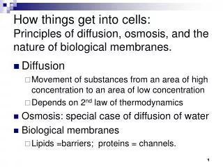
Diffusion MRI is an MR technique that can quantify the magnitude of H20 diffusivity within tissues Microstructural information ADC calculated from diffusion MR images is influenced by: Cellularity Tissue viscosity and temperature Diffusion Time Compartment Size (cell size and shape) Tortuosity of environment DTI is useful for directionality of diffusion restrictions CNS, MSK, Kidney DWI/ADC maps can be used to characterize many pathologies of the abdomen Liver and Kidney pathologies are most common abdominal diffusion MR studies Summary B.M. Ellingson, Ph.D., Dept. of Radiological Sciences, David Geffen School of Medicine at UCLA, 2010