Download
1 / 64
640 likes | 881 Vues
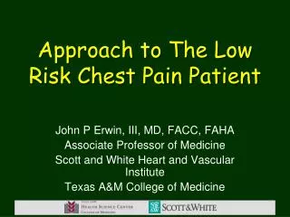
TRIAGE OF THE ED PATIENT COMPLAINING OF CHEST PAIN. David Plaut Snow, 2004. TRIAGE OF THE ED PATIENT COMPLAINING OF CHEST PAIN. 100%. ~4% AMI ND-ECG. AMI-DIAGNOSTIC ECG. AMI-NON DIAGNOSTIC ECG. NO AMI. 90%. Questionable Admissions 30%. Unstable angina, stable angina and

E N D
TRIAGE OF THE ED PATIENT COMPLAINING OF CHEST PAIN David Plaut Snow, 2004
TRIAGE OF THE ED PATIENT COMPLAINING OF CHEST PAIN 100% ~4% AMI ND-ECG AMI-DIAGNOSTIC ECG AMI-NON DIAGNOSTIC ECG NO AMI 90% Questionable Admissions 30% Unstable angina, stable angina and other acute coronary syndromes 30% Unnecessary Admissions 30% 0% 500,000 PATIENTS SENT HOME 5,000,000 PATIENTS ADMITTED CAP TODAY 1:51, 1994
Time to Presentation PERCENT OF PATIENTS ONSET TO PRESENTATION (HOURS) Note: 50 % present within 4 Hours (GISSI-3 STUDY POPULATION)
Reference Range lie on a continuuuuum TCK 0 ------------------------> 180 CK-MB 0 ------------------------> 5 Myo 0 ------------------------> 80 Age? Sex? Muscle mass? Genes?
cTn Reference Value. Normal Value for cTnI 0.0
Time TCK MB RI MYO cTnI <200 <5.0 <2.5 <80 <0.06 0 h 123 2.5 2.0 34 0.0 Case A A 40 yr old male with CP for 2 hours. His ECG was non-diagnostic.
Time TCK MB RI MYO cTnI <200 <5.0 <2.5 <80 <0.06 0 h 123 2.5 2.0 34 0.0 1 116 2.3 2.0 27 0.0 2 131 2.7 2.0 33 0.0 6 125 2.5 2.0 31 0.0 Case A A 40 yr old male with CP for 2 hours. His ECG was non-diagnostic. D’Costa et al. found a negative predictive value of 100% of Myo. at 2 hours. This was confirmed by Kircher and Montague.
Case B A 76 yr old male with a history of IHD and mild CHF. Presents with severe chest pain which did not diminish with nitroglycerin. cTnI Time MYO <0.06 <80 <0.06 66 0 h
Case B • A 76 yr old male with a history of IHD and mild CHF. Presents with severe chest pain which did not diminish with nitroglycerin. • TimeMYOcTnI • 0 h 66 <0.06 • 3 1470.47 • As many as 34% AMI present with a “normal” cardiac profile.
Case B • A 76 yr old male with a history of IHD and mild CHF. Presents with severe chest pain which did not diminish with nitroglycerin. • TimeMYOcTnI • 0 h 66 <0.06 • 3 1470.47 • 6 --- 1.30 • As many as 34% AMI present with a “normal” cardiac profile.
Time TCK MB RI MYO cTnI <200 <5.0 <2.5 <80 <0.06 0 h 817 29 3.5 82 54 1 756 24 3.2 82 44 12 241 4.0 1.6 43 21 Case C A 48 yr old male complained of CP after working in his field all morning. After trying Maalox he presented to the ED the following morning. Ladenson has found that cTnI remains detectable for as long as 15 days following an AMI.
Time TCK MB RI MYO cTnI <200 <5.0 <2.5 <80 <0.06 0 h 411 5.4 1.3 217 0.0 2 319 4.7 1.2 202 0.0 6 312 2.1 1.0 207 0.0 Case D A 64 yr old female with known chronic renal failure presents to the ED with “some pain in my chest.” Her EKG was non-diagnostic. Final diagnosis: Renal failure
Time TCK CK-MB RI Myo cTnI <200 <5.0 <2.5 <80 <0.06 0 h 32 -- -- 27 0.0 4 46 -- -- 67 1.1 Case E A 83 yr old female with intermittent chest discomfort is admitted to the ED at Huntington Hospital in Pasadena, CA.
Time TCK CK-MB RI Myo cTnI <200 <5.0 <2.5 <80 <0.06 0 h 32 -- -- 27 0.0 4 46 -- -- 67 1.1 9 56 -- -- 32 2.2 16 134 10.2 3.2 145 5.3 Case E A 83 yr old female with intermittent chest discomfort is admitted to the ED at Huntington Hospital in Pasadena, CA. Final diagnosis: AMI with extension
Questions Which marker(s)? When?
A 6 hour protocol for chest pain evaluation n = 292 (239 non-MI, 53 MI) • Sensitivity: 97.2%, specificity: 93% • The negative predictive value: 99.6% • “The six hour rule-out protocol is… accurate and efficacious.” Herren, BMJ 2001 Aug 18; 323:372 .
A 90 minute accelerated critical pathway for chest pain evaluation n = 1285 • All AMI’s were diagnosed within 90 min. • Negative predictive value: 100% • Ninety percent of patients with negative cardiac markers and a negative ECG at 90 minutes were discharged home Ng, S., Am J Cardiol 2001 Sept 15;88(6) 611-7
Evaluation of a 90 minute protocol n= 817 • Sensitivity: 96.9% • Negative predictive value: 99.6% • Addition of CK-MB did not improve the sensitivity or the NPV • Addition of a 3 hour draw did not improve sensitivity or the NPV McCord, Circulation.2001 Sept 25;104(13):1454-6
Suggested Protocol T0 Draw sample for cTn (and Myo?) If cTn is diagnostic discontinue order If cTn is not diagnostic Draw 2nd sample 2 - 3 hrs. later If cTn is diagnostic discontinue order If cTn is not diagnostic Draw 3d sample 2 - 3 hrs. later
TRIAGE OF ED PATIENTS COMPLAINING OF CHEST PAIN Unstable angina, stable angina and other acute coronary syndromes ~ 30% CAP TODAY 1:51, 1994
Unstable angina is a time bomb … A 68 yr old male with SOB, known chronic renal failure and acute renal insufficiency presents to the ED. His EKG was non-diagnostic. • TimecTnI • 0 h 0.36 • 9 0.35 • 33 0.32 Final diagnosis: Renal failure with CAD. Patient was discharged.
waiting to EXPLODE ! Three weeks later patient returned with severe chest pain and radiating left arm pain. • TimecTnI • 0 0.46 • 2 0.69 • 6 2.90
Serum cardiac troponin I values in unstable angina. • 74 patients with chest pain at rest, electrocardiographic evidence of myocardial ischemia, and normal values of CK-MB • Death or nonfatal myocardial infarction was more frequent in patients with elevated cTnI (27.7% vs 5.3%) than those with normal values. Ottani F Am Heart J 1999 Feb;137(2):284-91
cTnI to Predict Risk of Mortality in ACS Antman et al. NEJM 1996; 335:1342-9
TRIAGE OF ED PATIENTS COMPLAINING OF CHEST PAIN Unstable angina, stable angina and other acute coronary syndromes ~ 30% CAP TODAY 1:51, 1994
Total Cholesterol Distribution:CHD vs. Non-CHD Population Framingham Heart Study—26-Year Follow-up No CHD 35% of CHDoOccurs in people with TC <200 mg/dL CHD 150 300 250 200 Total Cholesterol (mg/dL) Adapted from Castelli. Atherosclerosis. 1996;124(suppl):S1-S9.
Questions: Why add another test? Why should it be hs-CRP?
Is there clinical evidence that hs-CRP, a marker of low grade vascular inflammation, predicts future coronary events?
3 2 1 0 < 0.055 0.056–0.114 0.115–0.210 ³ 0.211 hs-CRP and Risk of Future MI in Apparently Healthy Men P Trend <0.001 P<0.001 P<0.001 P=0.03 Relative Risk of MI 2 3 4 1 Quartile of hs-CRP (range, mg/dL) Ridker. N Engl J Med. 1997;336:973–979.
hs-CRP and Risk of Future Cardiovascular Events in Apparently Healthy Women P Trend <0.002 6 Any event 5 MI or stroke 4 RelativeRisk 3 2 1 0 1 2 3 4 < 0.15 0.15–0.37 0.37–0.73 > 0.73 Quartile of hs-CRP (range, mg/dL) Ridker. Circulation. 1998;98:731–733.
hs-CRP Adds to Predictive Value of TC:HDL Ratio in Determining Risk of First MI Relative Risk hs-CRP TC:HDL Ratio Ridker. Circulation. 1998;97:2007–2011.
Is there clinical evidence that the effect of hs-CRP on cardiovascular risk can be modified by preventive therapies?
hs-CRP, Aspirin, and Risks of Future Myocardial Infarction 4 3 Relative Risk Myocardial Infarction 2 1 Placebo Aspirin 0 4 3 2 1 Quartile of C-Reactive Protein Ridker PM, N Engl J Med 1997;336:973-9
What are the recommended guidelines for the use of hs-CRP assays?
Guidelines for Use of hs-CRP the writing group “recommends against screening the entire adult population for hs-CRP….” “it is reasonable to measure hs-CRP as an adjunct…to further assess absolute risk for CAD primary prevention.” Circulation 107 (Jan) 499, 2003
Relative Risk and Average hs-CRP hs-CRP < 1.0 mg/L Low 1.0 -- 3.0 Average >3.01 High
The Importance of the D-dimer Assay andIts Use in the Clinical SettingDavid Plaut
ThromboembolismIncidence & Mortality • DVT affects 2 million Americans per year • Without treatment, PE mortality~ 30% • With treatment of heparin or TPA, mortality is <2% • Only 15-25% of patients suspected of DVT/PE actually have DVT/PE.
Causes of Elevated D-dimer Atherosclerosis Trauma Hepatic disease DIC Infection Pregnancy Inflammation Age Cancer DVT Thrombolytic Rx PE
What is the importance of a negative D-dimer test? If D-Dimer is negative, then there are no clots being dissolved = no DVT or PE The value lies in the ability of d-dimer assays torule outthe Dx of DVT and PE
Clinical policy, College Emergency Physicians, 2003 Patient management recommendations Level A (high clinical certainty) None specified Ann. Emer. Med 41: 257, 2003
Clinical policy, College Emergency Physicians, 2003 Patient management recommendations Level B (moderate) Low pretest probability of PE use the following tests to exclude PE: 1. A negative quantitative d-dimer 2. A negative qualitative d dimer if Wells score 2 or less.
Clinical policy, College Emergency Physicians, 2003 Patient management recommendations Level C (low) Low pretest prob. of PE use the following tests to exclude PE: A negative quantitative d-dimer or a negative qualitative d dimer (when not used with Wells system)
Wells et al. criteria Suspected DVT 3.0 Alternate Dx is less likely than PE 3.0 Heart rate >100 1.5 Immobilized or surgery in last 4 wk 1.5 Previous DVT/PE 1.5 Hemoptysis 1.0 Malignancy (treated within is 6 mo.) 1.0 Wells, PS et al. Thromb Haemost. 83: 416, 2000
Wells score and probabilities for PE Score Probability 0 - 2 3.6% 3 - 6 20 >6 67
Use of D dimer to rule out DVT/PE Prevalence = 29% Sensitivity = 99.5 NPV = 99 Specificity = 41 n= 671 Am. J. Resp. Care 156: 492, 1997
Validity of D-dimer for DVT (Venography) Ten studies with 945 patients Sensitivity = 97% ( 89 – 100) NPV = 97 ( 92 – 100) Specificity = 54 ( 34 – 80) Brill-Edwards, P Thromb. Hemosta. 82: 688, 1999