Download
1 / 44
450 likes | 1.16k Vues
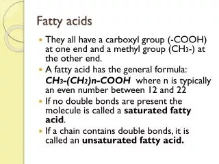
Synthesis and degradation of fatty acids. Zdeňka Klusáčková. Fatty acids (FA). mostly an even number of carbon atoms and linear chain. in esterified form as component of lipids. in unesterified form in plasma. binding to albumin. Groups of FA:. according to the chain length. <C 6.

E N D
Synthesis and degradation of fatty acids Zdeňka Klusáčková
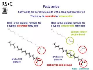
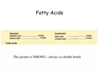
Fatty acids (FA) • mostly an even number of carbon atoms and linear chain • in esterified form as component of lipids • in unesterified form in plasma binding to albumin Groups of FA: • according to the chain length <C6 short-chain FA (SCFA) C6 – C12 medium-chain FA (MCFA) C12 – C20 long-chain FA (LCFA) >C20 very-long-chain FA (VLCFA) • according to the number of double bonds no double bond saturated FA (SAFA) one double bond monounsaturated FA (MUFA) more double bonds polyunsaturated FA (PUFA)
Triacylglycerols • main storage form of FA • acylglycerols with three acyl groups • stored mainly in adipose tissue
FA biosynthesis function: energy storage in the form of TAG • FA biosynthesis in the excess of energy (increased caloric intake) • acyl-CoA and glycerol-3-phosphate synthesis of TAG in liver • TAG incorporation into very low density lipoproteins (VLDL) • entry of VLDL into the blood circulation • TAG transport from the liver to other tissues via VLDL (especially skeletal muscle, adipose tissue)
FA biosynthesis • mainly in the liver, adipose tissue, mammary gland during lactation (always in excess calories) localization: • cell cytoplasm (up to C16) • endoplasmic reticulum, mitochondrion (elongation = chain extension) enzymes: • acetyl-CoA-carboxylase (HCO3- - source of CO2, biotin, ATP) • fatty acid synthase (NADPH + H+, pantothenic acid) primary substrate: • acetyl-CoA final product: • palmitate
FA biosynthesis • on the multienzyme complex – FA synthase • repeated extension of FA by two carbons in each cycle • to the chain length C16 (palmitate) • palmitate, a precursor of saturated and unsaturated FA: saturated FA (> C16) elongation systems desaturation systems unsaturated FA
Precursors for FA biosynthesis 1. Acetyl-CoA source: • oxidative decarboxylation of pyruvate (the main source of glucose) • degradation of FA, ketones, ketogenic amino acids • transport across the inner mitochondrial membrane as citrate 2. NADPH source: • pentose phosphate pathway (the main source) • theconversionofmalate to pyruvate • (NADP+-dependentmalatedehydrogenase - „malic enzyme”) • theconversionofisocitrate to α-ketoglutarate • (isocitratedehydrogenase)
FA biosynthesis Formation of malonyl-CoA HCO3- + ATP ADP + Pi enzyme-biotin enzyme-biotin-COO- biotinyl-enzyme carboxybiotinyl-enzyme 1 carboxylation of biotin 2 transfer of carboxyl group to acetyl-CoA acetyl-CoA formation of malonyl-CoA enzyme-biotin + enzyme – acetyl-CoA-carboxylase malonyl-CoA
FA biosynthesis Regulation at the level of ACC glucagon adrenaline cAMP insulin AMP protein kinase A AMP-dependent protein kinase A acetyl-CoA malonyl-CoA glucose citrate palmitate palmitoyl-CoA acetyl-CoA carboxylase
FA biosynthesis FA synthase
FA biosynthesis The course of FA biosynthesis acetyl-CoA malonyl-CoA CoASH CoASH acetyltransacylase malonyltransacylase transacylation acyl(acetyl)-malonyl- -enzyme complex
FA biosynthesis The course of FA biosynthesis 3-ketoacyl-synthase CO2 condensation acyl(acetyl)-malonyl-enzyme complex 3-ketoacyl-enzyme complex (acetacetyl-enzyme complex)
FA biosynthesis The course of FA biosynthesis NADP+ NADPH + H+ NADP+ NADPH + H+ H2O 3-ketoacyl-reductase 3-hydroxyacyl- dehydrase enoylreductase first reduction dehydration second reduction 3-ketoacyl-enzyme complex (acetoacetyl-enzyme complex) 3-hydroxyacyl-enzyme complex 2,3-unsaturated acyl-enzyme complex acyl-enzyme complex
FA biosynthesis Repetition of the cycle malonyl-CoA CoASH acyl-enzyme complex (palmitoyl-enzyme complex)
FA biosynthesis The release of palmitate thioesterase + H2O palmitate palmitoyl-enzyme complex
FA biosynthesis The fate of palmitate after FA biosynthesis acylglycerols cholesterol esters ATP + CoA AMP + PPi esterification palmitate palmitoyl-CoA acyl-CoA-synthetase elongation desaturation acyl-CoA
FA biosynthesis FA elongation 1. microsomal elongation system • in the endoplasmic reticulum • malonyl-CoA – the donor of the C2 units NADPH + H+ – the donor of the reducing equivalents • extension of saturated and unsaturated FA FA > C16 elongases (chain elongation) palmitic acid (C16) fatty acid synthase 2. mitochondrial elongation system • in mitochondria • acetyl-CoA – the donor of the C2 unit • not reverse β-oxidation
FA biosynthesis Microsomal extension of FA CoASH + CO2 + synthase acetyl-CoA malonyl-CoA 3-ketoacyl-CoA NADPH + H+ NADP+ H2O NADPH + H+ NADP+ hydratase reductase reductase 3-hydroxyacyl-CoA 2,3-unsaturated acyl-CoA acyl-CoA Example: CoASH + CO2 + palmitoyl-CoA malonyl-CoA NADPH + H+ NADP+ NADPH + H+ NADP+ H2O stearoyl-CoA
FA biosynthesis FA desaturation • in the endoplasmic reticulum • process requiring O2, NADH, cytochrome b5
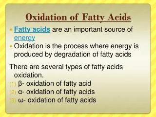
FA degradation function: major energy source (especially between meals, at night, in increased demand for energy intake – exercise) • release of FA from triacylglycerols in adipose tissue into the bloodstream • binding of FA to albumin in the bloodstream • transport to tissues • entry of FA into target cells activation to acyl-CoA • transfer of acyl-CoA via carnitine system into mitochondria β-oxidation Most important FA released from adipose tissue: • palmitic acid • oleic acid • stearic acid
FA degradation Mechanisms of FA degradation long-chain FA (LCFA, C12 – C20) mitochondrial β-oxidation unsaturated FA modified odd-chain-length FA mitochondrial β-oxidation peroxisomal β-oxidation very-long-chain FA (VLCFA, > C20) peroxisomal α-oxidation long-chain branched-chain FA FA with C10 or C12 ω-oxidation
FA degradation Mechanisms of FA degradation β-oxidation ω-oxidation α-oxidation
FA degradation β-oxidation • mainly in muscles localization: • mitochondrial matrix • peroxisome enzymes: • acyl CoA synthetase • carnitine palmitoyl transferase I, II; carnitine acylcarnitine translocase • dehydrogenase (FAD, NAD+), hydratase, thiolase substrate: • acyl-CoA final products: • acetyl-CoA • propionyl-CoA
FA degradation β-oxidation • repeated shortening of FA by two carbons in each cycle • cleavage of two carbon atoms in the form of acetyl-CoA • oxidation of acetyl-CoA to CO2 and H2O in the citric acid cycle complete oxidation of FA • generation of 8 molecules of acetyl-CoA from 1 molecule of palmitoyl-CoA • production of NADH, FADH2 reoxidation in the respiratory chain to form ATP PRODUCTION OF LARGE QUANTITY OF ATP
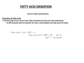
FA degradation Activation of FA fatty acid ATP acyl-CoA-synthetase acyl adenylate pyrophosphate (PPi) acyl-CoA-synthetase pyrophosphatase 2Pi acyl-CoA AMP fatty acid+ ATP + CoASH acyl-CoA + AMP + PPi PPi + H2O 2Pi
FA degradation The role of carnitine in the transport of FA into mitochondrion FA transfer across the inner mitochondrial membrane by carnitine and three enzymes: • carnitinepalmitoyltransferase I (CPT I) • acyl transfer to carnitine • carnitineacylcarnitinetranslocase • acylcarnitine transfer across • theinnermitochondrialmembrane • carnitinepalmitoyltransferase II (CPT II) • acyl transfer fromacylcarnitineback to CoA in themitochondrial matrix
FA degradation β-oxidation Steps of cycle: acyl-CoA • dehydrogenation • oxidation by FAD • creationofunsaturated acid acyl-CoA-dehydrogenase trans-Δ2-enoyl-CoA • hydration • additionofwater on theβ-carbon atom • creationofβ-hydroxyacid enoyl-CoA-hydratase L-β-hydroxyacyl-CoA L-β-hydroxyacyl-CoA- • dehydrogenation • oxidation by NAD+ • creationofβ-oxoacid -dehydrogenase β-ketoacyl-CoA • cleavageatthe presence ofCoA • formationof acetyl-CoA • formationof acyl-CoA (twocarbonsshorter) β-ketoacyl-CoA-thiolase acyl-CoA acetyl-CoA
FA degradation Oxidation of unsaturated FA • the most common unsaturated FA in the diet: linoleoyl-CoA cis Δ9, cis-Δ12 oleic acid,linoleic acid 3 rounds of β-oxidation 3 acetyl-CoA • degradation of unsaturated FA • by β-oxidation to a double bond cis-Δ3, cis-Δ6 enoyl-CoA-isomerase • conversion of cis-isomer of FA • by specific isomerase to trans-isomer trans-Δ2, cis-Δ6 β-oxidation 1 acetyl-CoA • continuation of β-oxidation • to the next double bond cis-Δ4 acyl-CoA-dehydrogenase • formation of double bond between C2 and C3 by dehydrogenation trans-Δ2, cis-Δ4 NADPH + H+ • elimination of double bond between C4 and C5 by reduction dienoyl-CoA-reductase NADP+ trans-Δ3 enoyl-CoA-isomerase • intramolecular transfer of double bond trans-Δ2 4 rounds of β-oxidation • continuation of β-oxidation 5 acetyl-CoA
FA degradation Oxidation of odd-chain FA propionyl-CoA • shortening of FA to C5 stopping of β-oxidation HCO3- + ATP propionyl-CoA carboxylase (biotin) ADP + Pi • formation of acetyl-CoA and propionyl-CoA D-methylmalonyl-CoA • carboxylation of propionyl-CoA methylmalonyl-CoA racemase • epimerization of D-form into L-form L-methylmalonyl-CoA • intramolecular rearrangement to form succinyl-CoA methylmalonyl-CoA mutase (B12) • entry of succinyl-CoA into the citric acid cycle succinyl-CoA
FA degradation Peroxisomal oxidation of FA A)very-long-chain FA (VLCFA, > C20) • transport of acyl-CoA into the peroxisome without carnitine Differences between β-oxidation in the mitochondrion and peroxisome: 1. step – dehydrogenation by FAD mitochondrion: electronsfrom FADH2 are delivered to therespiratorychain wherethey are transferred to O2 to form H2O and ATP peroxisome: electrons from FADH2 aredelivered to O2 to form H2O2,which is degraded by catalase to H2O and O2 3. step – dehydrogenation by NAD+ mitochondrion: reoxidation of NADH in the respiratory chain peroxisome: reoxidation of NADH is not possible, export to the cytosol or the mitochondrion
FA degradation Peroxisomal oxidation of FA Differences between β-oxidation in the mitochondrion and peroxisome: 4. step – cleavage at the presence of CoA acetyl-CoA mitochondrion: metabolization in the citric acid cycle peroxisome: export to the cytosol, to the mitochondrion (oxidation) a precursor for the synthesis of cholesterol and bile acids a precursor for the synthesis of fatty acids of phospholipids
FA degradation Peroxisomal oxidation of FA B)long-chain branched-chain FA • blocking of β-oxidation by the alcyl group at Cβ • α-oxidation • hydroxylation at Cα • cleavage of the original carboxyl group as CO2 • methyl group is in the position α • shortening of FA to 8 carbons • transfer of FA in the form of acylcarnitine into the mitochondrion • complete of β-oxidation in the mitochondrion
Refsum's disease • rare autosomal recessive hereditary disease • phytanic acid a product of metabolism of phytol (part of chlorophyll) in milk and animal fats • decreased activity of peroxisomal α-hydroxylase accumulation of phytanic acid (in tissues of nervous system and serum) • ataxia, night blindness, hearing loss, skin changes etc.
FA degradation ω-oxidation of FA • minor pathway of FA oxidation • in the endoplasmatic reticulum • repeated oxidation of ω-carbon -CH3 -CH2OH -COOH • formation of dicarboxylic acid • entry of dicarboxylic acid into β-oxidation • reduction of FA to adipic acid (C6) or suberic acid (C8) excreted in the urine
FA degradation Regulation of β-oxidation A) by energy demands of cell by the level of ATP and NADH: FA can not be oxidized faster than NADH andFADH2 arereoxidized in the respiratory chain B) via carnitine palmitoyl transferase I (CPT I) CPT I is inhibited by malonyl-CoA, which is generated in the synthesis of FA by acetyl-CoA carboxylase (ACC) active FA synthesis inhibition of β-oxidation acetyl-CoA malonyl-CoA CPT I β-oxidation ACC
Ketone bodies Ketogenesis • in the liver localization: • mitochondrial matrix substrate: • acetyl-CoA products: • acetone • acetoacetate • D-β-hydroxybutyrate conditions: • in excess of acetyl-CoA function: • energy substrates for extrahepatic tissues
Ketone bodies Ketogenesis
Ketone bodies Ketogenesis acetoacetate • spontaneous decarboxylation to acetone • conversion to D-β-hydroxybutyrate • by D-β-hydroxybutyrate dehydrogenase waste product (lung, urine) energy substrates for extrahepatic tissues
Ketone bodies Utilization of ketone bodies • water-soluble FA equivalents • energy source for extrahepatic tissues (especially heart and skeletal muscle) • in starvation - the main source of energy for the brain energy citric acid cycle production
Ketone bodies Production, utilization and excretion of ketone bodies acetyl-CoA • oxidation in the citric acid cycle (liver) • conversion to ketone bodies (liver - mitochondrion) • release of ketone bodies into blood • transport to tissues
Ketone bodies Ketogenesis increased ketogenesis: lipolysis • starvation • prolonged exercise • diabetes mellitus FA in plasma • high-fat diet • low-carbohydrate diet β-oxidation utilization of ketone bodies as an energy source (skeletal muscle, intestinal mucose, adipocytes, brain, heart etc.) excess of acetyl-CoA to spare of glucose and muscle proteins ketogenesis
Bibliography and sources Devlin, T. M. Textbook of biochemistry: with clinical correlations. 6th edition. Wiley-Liss, 2006. Marks, A.; Lieberman, M. Marks' basic medical biochemistry: a clinical approach. 3rd edition. Lippincott Williams & Wilkins, 2009. Matouš a kol. Základy lékařské chemie a biochemie. Galén, 2010. Meisenberg, G.; Simmons, W. H. Principles of medical biochemistry. 2nd edition. Elsevier, 2006. Murray et al. Harper's Biochemistry. 25th edition. Appleton & Lange, 2000. http://www.hindawi.com/journals/jobes/2011/482021/fig2/