Download
1 / 70
700 likes | 1.1k Vues
ENDOCRINE SYSTEM ENDOKRIEN SISTEEM. Dr Resia Pretorius Hoof: Afdeling Histologie, Department Anatomie Kamer 6-27 Departement Anatomie Tel: 319 2533. Division / Indeling. Pituitary / hipofese Hipothalamus / hypothalamus Thyroid / skildklier Parathyroid/ byskildklier Thymys / timus

E N D
ENDOCRINE SYSTEMENDOKRIEN SISTEEM Dr Resia Pretorius Hoof: Afdeling Histologie, Department Anatomie Kamer 6-27 Departement Anatomie Tel: 319 2533
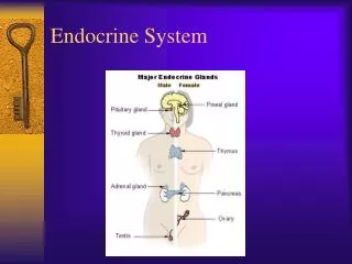
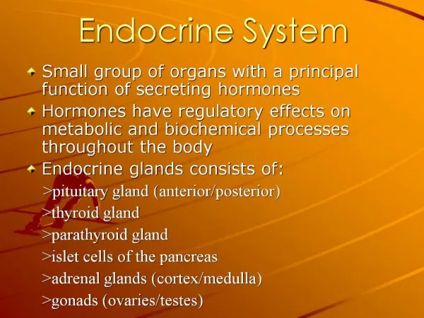
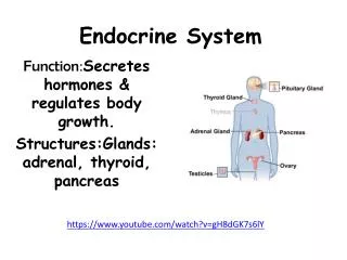
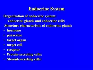
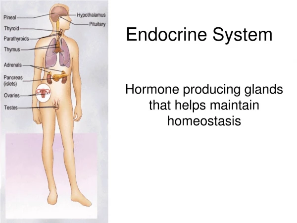
Division/ Indeling • Pituitary / hipofese • Hipothalamus / hypothalamus • Thyroid / skildklier • Parathyroid/byskildklier • Thymys / timus • pineal/ pineaalklier • kidney/ nier • pancreas/pankreas • adrenals / bynier of adrenaalklier • ovary/ovaria • testis
Pituitary/hipofese • Underneath the base of the brain • attached via stem = infundibulum • Gland is split into anterior and posterior area (adult absent) • dura mater surrounds gland • rest on sfenoid bone - cells of turcica • 4 areas
Pars anterior (pars distalis, anterior lobe) • pars tuberalis • pars intermedia • pars posterior (pars nervosa, posterior lobe)
Pars tuberalis Pars distalis pars intermedia Anterior lob • Adenohipofese • Neurohipofese Pars nervosa Infundibulum Emmenentia medialis Posterior lob
Embryonic development of Pituitary gland • Derived from an ectodermal outpocketing of the stomodeum (Rathke's Pouch) and in part from the floor of the diencephalon (infundibulum).
Embryological development • stomodeum (Rathke's Pouch) • diencephalon (infundibulum).
Embryological development • Adenohypophysis (anterior lobe) is derived from Rathke's Pouch and the neurohypophysis (pars nervosa) is derived from the infundibulum
The larger portion, (adenohypophysis), is toward the top. • Left - superior aspect with the stalk coming • from the hypothalamus entering it. • Inferior - right. • The posterior pituitary (neurohypophysis) is the smaller portion at the bottom.
Pituitary (fine structure)Hipofese (mikroskopiese struktuur) • The adenohypophysis is at the right • The neurohypophysis is at the left.
Adenohypophysis: cells • ACIDOPHILS (35%) (pink) • growth hormone (GH) and prolactin (PRL) • BASOPHILS (15%)(Dark purple) • corticotrophin (ACTH), • thyroid stimulating hormone (TSH), and gonadotrophins • follicle stimulating hormone-luteinizing hormone (FSH and LH) • CHROMATOPHOBES (chromofobe)(50%) (Pale staining) • few cytoplasmic granules, but may have secretory activity.
Pars distalis(anterior lobe/adenohipofese) • 75% of the anterior lobe • cells form branched chords • between cells fenestrated capillaries • Cells: • CHROMOPHOBES(chromatofobe) resting AND CHROMOPHILS (hormone sectreting) • = asidophils and basophils
Pars tuberalis • Collar of cells surrounding infundibulum • kraag selle • Upwards lengthening of pars distalis • rich blood supply • Endocrine cells in groups (cuboidal) koorde (kubies) • Nests of flattened cells andsmall follicles (nessies plat selle en klein follikels) • function: unknown
Pars intermedia • Not well developed • residual lumen of Rathke’s pouch oorblyfsel van Rathke se sak • Colloid filled cysts kolloid • endocrine cells
Neurohypophysis • Pars nervosa • infundibulum • Emmenentia medialis
Neurohipofese • resembles neural issue, with glial cells, nerve fibers, nerve endings, and intra-axonal neurosecretory granules. • hormones transported into the intra-axonal neurosecretory granules where they are released.
Pars nervosa (Neurohipofese en posterior lob) • Neuron cell bodies • Nerve fibers • Pituicytes (neuroglia; astrocytes) • Nerve endings • Herring bodies
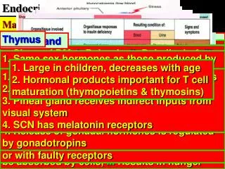
PINEAL GLAND/PINEAALKLIER • The pineal gland was called the "third eye" by ancient people. • center for the production of the hormone melatonin. • Melatonin is implicated in a wide range of human activities. • It regulates daily body rhythms, most notably the day/night cycle (circadian rhythms).
Claims that the hormone slows the aging • process (a defense against free radicals) • prevents jet lag, is implicated in seasonal affective disorder, coordinates fertility, • allows for deep restful sleep patterns.
The pineal gland calcifies with age and melatonin production correspondingly decreases. • This decline in melatonin has been suggested to be a trigger for the aging process. • pineal gland is a center for navigation in birds
Pineaalklier • Pia mater om klier = septa • lobules • Pinealosiete ( irregular) vertakte prosesse • neuroglia (ondersteunend)
Thyroid • Structural unit = follicle; connective tissue capsule - lobes • follicular cells or principal cells spoed van metabolisme • parafollicular cells or C cells verlaag bloedkalsium • colloid (inactive storage form of thyroglobulin) • fenestrated capillaries and reticular network
Thyroid papillary carcinoma • The most favorite site of metastasis • is to local lymph nodes in the neck.
Thyroid medullary carcinoma • At the center and to the right is a medullary carcinoma of thyroid. • These neoplasms are derived from the thyroid "C" cells and, therefore, have neuroendocrine features such as ecretion of calcitonin.
Parathyroids/ byskildklier • Endocrine cells denely packed into chords • principal cells/ klein selle; glikogeen; kalsiummetabolisme • Oxyphil cells/ oksifiele • Fenestrated capillaries
Adrenals/ bynier • Cortex (90%) mesoderm - steroid secretion portion • medulla (10%)ectoderm - catecholamine secretion • rich blood supply - fenestrated capillaries • reticular network
Cortex • Zona glomerulosa (15%) onder kapsel • klein onreëlmatige groepies; kapillêre tussenin • Zona fasciculata (78%) • reguit koorde selle; veelhoekig; kapillêre tussen koorde • Zona reticularis (7%) network dark nuclei
Medulla • Endocrine cells, connective tissue, blood vessels, nerves • cells in groups and chords • chromaffin cells (chroomneerslag - kleur bruin)
Zona glomerulosa and Zona fasciculata • Gigher magnification of the zona glomerulosa and the Zona fasciculata is shown in this photo. Note the many droplets • seen in the zona fasciculata cells.
Hormone producing cells in organs • Gonads • liver • kidney • pancreas • thymus