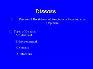
Hirschsprung Disease
390 likes | 1.43k Vues
Alquds university. Hirschsprung Disease. Prepared by Maha Hmeidan /Nahal RN MSN. Hirschprung’s Disease. More than 50 years old since the discovery of the cause and the treatment for Hirschprung disease. Incidence : 1 in 5000 live birth 80 % of patients are males.

Hirschsprung Disease
E N D
Presentation Transcript
Alquds university Hirschsprung Disease Prepared by Maha Hmeidan /Nahal RN MSN
Hirschprung’s Disease • More than 50 years old since the discovery of the cause and the treatment for Hirschprung disease. • Incidence : 1 in 5000 live birth • 80 % of patients are males
Hirschsprung Disease Causes • Association with inheritance in chromosome 10 in some patients. • Autosomal Recessive • Autosomal dominant in totally agangloinic bowel. • Common in Down syndrome
Hirschsprung Disease • Aganglionosis is restrictedto the rectum and sigmoid colon in 75% of patients . • extends moreproximally in 15-20% . • and affects the entire colon and a variablelength of ileum in 8%. • Rarely, ganglion cells are absent frommost of the gastrointestinaltract.
Why Hirschsprung Disease cause constipation • This absence of normal parasympathatic innervation prevents gut peristalsis, leading to functional constipation. • The proximal colon hypertrophied by trying to overcome functional obstruction. • Transitional zone exists between normal and abnormal aganglionic intestine.
Hirschsprung Disease • This results in failure of the internal sphincter to relax with rectal distention. • Acetylcholine concentrations in aganglionic segments are threefold lower than in ganglionic segments.
Clinical Manifestations / diagnosis • Only 15% are diagnosed in the first month of life, but two thirds are in the first 3 months. Cases beyond 5 years of age usually have ultra- short segment disease.
Clinical diagnosis Symptoms within the first week of life include • failure to pass meconium within 48 hours,, • reluctance to feed ,bilious vomiting, abdominal distention • irritability or frowning appearance. • Constipation. sometimes Small watery stool • Anemia. • Delayed Growth. • Fever due to infection (enterocolitis)
Clinical diagnosis • Explosive liquid stools, fever, and severe prostration are indicative of enterocolitis. • Enterocolitis is rare (10%) in the first month but rises to 33% in the second and third months. • Recall that diarrhea may be a late sign.
Diagnosis • Rectal exam when the rectal vault is found devoid of stool and the anal canal feels narrow with increased tone. • Abdominal x-ray. • Barium enema. • Manometry. a test that measures the movement and strength of the rectal and anal sphincter muscles. • Biopsy.
Abdominal X-raymultiple dilated loops of bowel and a low bowel obstruction.
Diagnosis • Barium enema - A contrast agent such as barium) is given into the rectum in order to coat the inside of organs so that they will show up on an X-ray. • ( narrow segment and dilated proximal portion). Late films will show retention of barium at 24- 48 hours.
Anorectal –manometry • This determines whether normal reflexes involving the rectum and the anus are present. Used only in older children, the test can be performed at the bedside. • A small balloon is inflated inside the rectum.
Rectal biopsy • is the most reliable diagnostic measure. Absence of ganglion cells in an infant is not conclusive because they may be small and monopolar.
The acetylcholinesterase stained section illustrates the increase in positively (darkly) staining fibers within the lamina propria and muscularis propria which, in the absence of ganglion cells, is diagnostic of Hirschsprung disease.
Complicatons ofHirschsprung`s disease include • intestinal perforation (particularly at the appendix) • Enterocolitis. • water intoxication, results from the use of tap water enemas.. • there may be hypertonic dehydration from saline enemas • malnutrition, failure to thrive, and anemia.
Treatment Surgical (Pull-through Surgery) • It involves taking out the diseased part of the intestine that doesn't work and connecting the healthy part that's left to the anus. After surgery, the child will recover well and his intestines will work normally. • Often, the surgery can be done right after the diagnosis.
Colostomy and Ileostomy • However, children who have been very sick may first need ostomy to help the child to restore his health before having the pull-through surgery. Some doctors do ostomy in every child before doing the pull-through. • In an ostomy, the doctor takes out the diseased part of the intestine, then cuts a small hole called stoma then doctor connects the top part of the intestine to the stoma. Stool leaves the body through the stoma while the bottom part of the intestine heals. • Stool goes into the stoma bag that is attached to the skin around the stoma.
Colostomy and Ileostomy • If surgery entailed removal of the entire large intestine, the small intestine are connected to the stoma, the surgery is called an ileostomy. • If part of the large intestine is left, then it will be connected to the stoma, the surgery is called a colostomy • Later on, the pull-through surgery is performed, the intestines are disconnected from the stoma and attach just above the anus and the stoma isn't needed any more. In this case either the stoma is sewed up during surgery or sewed later for about 6 weeks to make sure that the pull-through surgery is working well
Operative photograph showing a sleeve rectal mucosectomy being performed down to the anus.
Gross photograph (of another patient) of a distal colonic segment resected for Hirschsprung disease. Note the dilated, proximal portion separated from the constricted distal portion by a transition zone.
Post-operative Complications • Early : Anastomotic leak Infection • Late : Obstruction Enterocolitis Incontinence
Hospital care • The nurses can teach the family and the child how to care for a stoma and can talk to you about your worries. • After a pull-through surgery, 9 out of 10 children pass stool normally. • Some children may have diarrhea for a while, and babies may develop a sever diaper rash, eventually the stool will become more solid and the child will need to go to the bathroom less often. • Toilet training may be delayed, as the child learns how to use the bottom muscles only after pull-through surgery. • Older children might stain their underwear for a while after the surgery. It is not their fault. They can't control this problem, but it improves with time.
Diet and Nutrition • Drinking plenty of liquids is important after surgery for HD to make sure his body gets enough fluids. • An infant who has long-segment disease requiring an ileostomy may need special tube feedings to make up for what is lost. • Eating high-fiber foods like cereal and bran muffins can help reduce constipation and diarrhea.
Infections (enterocolitis). • Enterocolitis can be life threatening for a child with Hirschsprung's disease, It can happen before or after surgery, so watch for the serious signs and call your doctor immediately if they occur. • Serious signs as: • fever, swollen abdomen, vomiting and diarrhea, bleeding from the rectum, sluggishness and drowsiness • In the case of enterocolitis, the child should be admitted to the hospital: • In the hospital, an intravenous (I.V.) line may be needed to keep body fluids up and to deliver antibiotics to fight the infection.
The large intestine will be rinsed regularly with a mild salt water solution until all remaining stool has been removed. The rinse may also contain antibiotics to kill bacteria. • When the child recovers from infection, then surgery is advised, and colostomy or ileostomy is done before the child leaves the hospital.
Having healthy babies Best wishes Maha Hmeidan