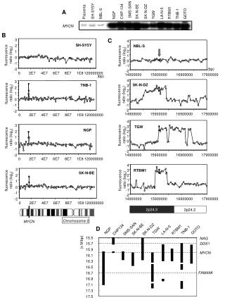
PLACENTA
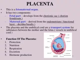
PLACENTA. This is a fetomaternal organ. It has two components: Fetal part – develops from the chorionic sac ( chorion frondosum ) Maternal part – derived from the endometrium ( functional layer – decidua basalis )

PLACENTA
E N D
Presentation Transcript
PLACENTA • This is a fetomaternal organ. • It has two components: • Fetal part – develops from the chorionic sac ( chorion frondosum ) • Maternal part – derived from the endometrium ( functional layer – decidua basalis ) • The placenta and the umbilical cord are a transport system for substances between the mother and the fetus.( vessels in umbilical cord ) • Function Of The Placenta: • Protection • Nutrition • Respiration • Excretion • Hormone production
Further Development of Chorionic Villi Early in the 3rd week, mesenchyme growth into the primary villi forming a core of mesenchymal tissue. Thus the Secondary Chorionic Villi are formed over the entire surface of the chorionic sac. Some mesenchymal cells in the secondary villi differentiate into capillaries and blood cells forming the Tertiary Chorionic Villi. The capillaries in the villi fuse to form arteriocapillary networks.
The previous formed arteriocapillary networks become connected with the embryonic heart through vessels which are formed in the mesenchyme of the chorion and connecting stalk. By the end of the 3rd week, embryonic blood begins to flow through the capillaries in the chorionic villi. Oxygen & nutrients in the maternal blood in the intervillous space diffuse through the walls of the villi and enter the embryo’s blood. Carbon dioxide & waste products diffuse from blood in the fetal capillaries through the wall of the chorionic villi into the maternal blood.
DECIDUA • This is the endometrium of the gravid (pregnant) uterus. • It has four parts: • Decidua basalis: it forms the maternal part of the placenta • Decidua capsularis: it covers the conceptus • Decidua parietalis: the rest of the endometrium • Decidua reflexa: • Junction between capsularis & parietalis.
DEVELOPMENT OF PLACENTA • Until the beginning of the 8th week, the entire chorionic sac is covered with villi. • After that, as the sac grows, only the part that is associated with Decidua basalis retain its villi. • Villi of Decidua capsularis compressed by the developing sac. • Thus, two types of chorion are formed: • Chorion frondosum (villous chorion) • Chorion laeve – bare (smooth) chorion • About 18 weeks old, it covers 15-30% of the decidua and weights about 1\ 6 of fetus
DEVELOPMENT OF PLACENTA • The villous chorion ( increase in number, enlarge and branch ) will form the fetal part of the placenta. • The decidua basalis will form the maternal part of the placenta. • The placenta will grow rapidly. • By the end of the 4th month, the decidua basalis is almost entirely replaced by the fetal part of the placenta.
FULL-TERM PLACENTA • Cotyledons –about 15 to 20 slightly bulging villous areas. Their surface is covered by shreds of decidua basalis from the uterine wall. • After birth, the placenta is always inspeced for missing cotyledons. Cotyledons remaining attached to the uterine wall after birth may cause severe bleeding. • Grooves – formerly occupied by placental septa • The fetal part of placenta; fetal membranes called developmental adnexa • Placenta;fetal membranes which are expelled are called afterbirth or secundina Maternal side
FULL-TERM PLACENTA( Discoid shape -500- 600 gm- Diameter 15-20 cm – Thicknessof 2-3 cm) • Fetal surface: • This side is smooth and shiny. It is covered by amnion. • The umbilical cord is attached close to the center of the placenta. • The umbilical vessels radiate from the umbilical cord. • They branch on the fetal surface to form chorionic vessels. • They enter the chorionic villi to form arteriocapillary-venous system. Fetal side
PLACENTAL MEMBRANE knot –syncytiotrophoblast –Toward end of pregnancy – phagocytic cells • This is a composite structure that consists of the extrafetal tissues separating the fetal blood from the maternal blood. • It has four layers: • Syncytiotrophoblast • Cytotrophoblast • Connective tissue of villus • Endothelium of fetal capillaries • After the 20th week, the cytotrophoblastic cells disappear and the placental membrane consists only of three layers.
TRANSFER ACROSS THE PLACENTAL MEMBRANE Viruses: measles;poliomyelitis Microorganism: treponema pallidum of syphilis ; T.g which produce destructive change in the eye; brain . IgG( gamma globulin) , IgS;IgM ( immunoglobulin S;M )
Placental endocrine synthesis • The syncytiotrophoblast synthesizes protein &steroid hormones • The protein homones • 1- human chorionic gonadotropin • 2- h.c. somatomammotropin • 3-h.c. thyrotropin • 4-h.c. corticotropin • The steroid hormones • Progesterone & Estrogens
Third trimester bleeding is the common sign of these anomalies
When villi persist on the entire surface of the chorionic sac ,a thin layer of placenta attaches to a large area of the uterus …… it is a membranous placenta.
FULL-TERM UMBILICAL CORD • Usually it is attached near the center of the fetal surface of placenta. • Length: about 50 cm • Diameter: 1-2 cm • Contains two arteries and one vein, surrounded by mucoid connective tissue (Wharton jelly) • The vessels are longer than the cord and may have loops (false knots).