Lecture 17
Lecture 17. DNA Replication – Process Chapter 30.1-30.4. DNA Replication. Forms of DNA Helices. Replication of parent DNA can theoretically generate 3 possible conformations of daughter DNA. Template DNA (parent DNA). S Phase. S Phase. M Phase. M Phase. DNA Replication.

Lecture 17
E N D
Presentation Transcript
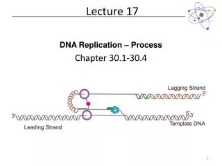
Lecture 17 DNA Replication – Process Chapter 30.1-30.4
DNA Replication Forms of DNA Helices Replication of parent DNA can theoretically generate 3 possible conformations of daughter DNA Template DNA (parent DNA) S Phase S Phase M Phase M Phase
DNA Replication Forms of DNA Helices In 1958, Meselson and Stahl designed an experiment to determine how DNA replication worked… http://palyapbio.edu.glogster.com/meselsonstahlcc
DNA Replication Forms of DNA Helices • So, shortly after Watson and Crick determined the structure of DNA (with err, “borrowed” data from Rosalind Franklin), Meselson and Stahl determined that DNA replication was semi-conservative. • Side note: People knew about the results of their experiments for months. Finally a colleague had to lock them in a room with cots and typewriters in order to get them to write the paper detailing their results. And you thought you procrastinated on lab reports! • But what does this mode of replication imply? • There must be a template strand • Did one parent DNA strand serve as a template or both? • Did DNA replication occur in both directions of the chromosome? • How was the DNA opened for replication? • What was responsible for the replication reaction?
DNA Replication Forms of DNA Helices How do bacteria go about replicating DNA? Linearize DNA for replication? Keep DNA circular? The experiment: Grow cells in 3H-thymidine enhanced media
DNA Replication Forms of DNA Helices Where does replication start and is it uni- or bidirectional? Heavy staining on both sides of the replication eye indicates that replication is almost always bidirectional in bacteria. Bacterial replication also starts at the same spot indicating a single origin of replication
Dealing with DNA Superstructure Forms of DNA Helices Parent DNA must be unwound at the replication fork For this to occur at biological rates (1000 nt per second for E. coli), genomic DNA must twist at a rate of 100 rps. What enzyme is capable of modifying DNA topology? Topoisomerase (DNA Gyrase) E. coli’s genome is circular, so this is not possible!
Semi-discontinuous Replication Forms of DNA Helices DNA’s 2 strands are replicated at the same time DNA Polymerase synthesize DNA in the 5’ 3’ direction 3’ 5’ 5’ So how does the other strand elongate? The ‘Lagging Strand’ is synthesized as small fragments (1000 – 2000 nuclotides in bacteria or 100-200 nt in eukaryotes) called Okazaki fragments. Okazaki fragments are combined by a DNA Ligase after the RNA primers are cut out by the 5’-3’ exonuclease activity of DNA PolI
DNA Replication Forms of DNA Helices How will a dNTP be added to the existing chain? How might the reaction be activated?
Requirement for priming Forms of DNA Helices This model of DNA replication requires a 3’ OH for strand elongation 5’ 3’ 5’ Analysis of Okazaki fragments indicated that the 5’ end was always composed of RNA 160 nucleotides in length These will have to be replaced at some point Nucleophile
Polymerization Reaction Forms of DNA Helices http://wiki.cstl.semo.edu/agathman/GetFile.aspx?File=DNA%20polymerase/nucleophilic.gif
Enzyme Requirements for Replication Forms of DNA Helices • DNA Polymerase • Catalyze the DNA chain elongation using the ‘parent’ strand as a template • RNA Primer Synthesis • RNA Polymerase • 460 kDa (E. coli) • catalyzes primer synthesis for leading strand only • Primase • 60 kDa • Responsible for priming Okazaki fragment • Works synergistically with RNA Polymerase to prime leading strand • Topoisomerase (Gyrase) • Unwind supercoiled template DNA • Energy dependent process • Ligase • Join Okazaki fragments
Formation of the Replication Fork Forms of DNA Helices • 4 DnaA proteins bind to the oriC region (recognizes 9 nucleotide segments) • Additional DnaA monomers bind forming a histone like complex of tightly wound DNA • Localized melting of a 13bp repeats • DnaB (6 subunit helicase) binds and unwinds the DNA • Topoisomerase functions downstream to relieve stress • Single Strand DNA binding proteins prevent the ssDNA from reannealing
Formation of the Replication Fork Forms of DNA Helices 178 Å DnaA helical filament – 8 monomers per turn • ATP-bound DnaA molecules sit on 3 of the 5 DnaA boxes of the origin. • They recruit additional DnaA molecules to occupy the remaining boxes • This introduces positive supercoiling into the DNA, causing the DNA to unwind (It is negatively supercoiled, right? Topoisomerase says “Oh, yeah.”) • The ATPase domain of DnaA may actively unwind DNA as a function of ATP hydrolysis • 4) The oriC-DnaA complex recruits DnaB-DnaChexamers to actively separate the DNA helix (DnaB is a helicase, DnaC is a loading protein that places DnaB onto the strand)
The Big Picture (in E.coli) Forms of DNA Helices Topoisomerase: relieves topological strain Helicase - unwinds dsDNA at replication fork SSB: keeps ssDNA from reannealing Primase: synthesizes RNA primers for lagging strand Pol III elongates lagging strand until strained. It then releases the template and rebinds at a new primed location. DNA Polymerase III : elongate primed DNA from 5’3’ 3’5’ exonuclease activity limits errors Pol I: closes any gaps in lagging strand and replaces RNA primer Ligase: seals off backbone nicks
What do we need in a DNA Polymerase? Forms of DNA Helices • Template DNA binding site • Single Strand or Double Strand? • Remember DnaA/DnaB/DnaC? • Room for growing dsDNA • dNTP binding site • Mechanism to differentiate between the 4 possible dNTPs • Self Correcting? • Viral Polymerase vs. Human Polymerase
DNA Polymerase Structures Forms of DNA Helices • Arthur Kornberg discovered the first DNA polymerase isolated from Escherichia coli in 1956 • This was called DNA Polymerase I (DNA PolI) • The structure of this enzyme was solved by Tom Steitz in 1987 (at least part of it was…) • PDB ID Code: 1DPI • The structure of the Thermusaquaticus DNA PolI was also discovered by Steitz in 1996 • PDB ID Code: 1TAQ • For today’s lectures, we’ll use the following structures: • 1QSS: ddGTP-trapped Closed Ternary Complex of the Large Fragment of DNA PolI from T. aquaticus • 1KLN: DNA PolIKlenow Fragment Mutant/DNA Complex
E. coli DNA Polymerase I Forms of DNA Helices • Pol I • 3’ 5’ and 5’ 3’exonuclease activity • Small N-terminal domain contains the 5’3’ exonuclease activity • Completely independent from active site or 3’ 5 site • Thumb • Guides newly formed DNA • Responsible for processivity • Fingers • Contains dNTP binding site • Close in on dNTP/Growing Strand once successful Watson-Crick base pairing made • Palm Domain • ~22 Å x 30 Å • Ideal shape to bind B-DNA • Lined with basic amino acids
3’ 5’ Exonuclease Forms of DNA Helices The structure of E. coli Pol I has been solved with DNA bound in the 3’5’ exonuclease active site Growing strand peels away from active site The base pair closest to the polymerization active site is significantly weakened Exonuclease site (Zn2+ activated mechanism) Which bond will be cleaved? NEED 3’-hydroxyl for elongation
Model for Proofreading in DNA Polymerases Forms of DNA Helices • dNTPs are in rapid equilibrium at active site • When reaction occurs, proper base pair will form a strong tight bonding pair • When an error occurs, the base pair is not as tight, resulting in increased favorability for the growing chain to be positioned at the 3’5’ exonuclease site • A-form DNA of DNA @ polymerization active site makes this process easier (less tightly wrapped and less stable helix)
3’->5’ Exonuclease Mechanism Forms of DNA Helices The structure of E. coli Pol I has been solved with DNA bound in the 3’5’ exonuclease site Growing strand peels away from active site Tyr 423 activates a water molecule which then serves as a nucleophile to begin the cleavage of the base from the strand
3’->5’ Exonuclease Mechanism Forms of DNA Helices Tyr 423 activates a water molecule which then serves as a nucleophile to begin the cleavage of the base from the strand Freemont et al. (1988). Cocrystal structure of an editing complex of Klenow fragment with DNA. PNAS 85: 8924-8928.
Taq DNA Polymerase Forms of DNA Helices Two domains were identified: Polymerase Domain Exonuclease Domain N-terminal domain contains 5’ 3’ exonuclease C-terminal domain contains polymerase activity
Taq DNA Polymerase Forms of DNA Helices • Notice the same Finger and Thumb domains as E. coli DNA PolI • Same function, same domains • Evidence of evolutionary conservation of function • No 3’->5’ exonuclease domain though…
E. coli and Taq DNA Polymerase Superposition Forms of DNA Helices • Notice the same Finger and Thumb domains as E. coli DNA PolI • Same function, same domains • Evidence of evolutionary conservation of function • No 3’->5’ exonuclease domain though…
Taq DNA Polymerase Forms of DNA Helices
Taq DNA Polymerase Forms of DNA Helices
Taq DNA Polymerase Forms of DNA Helices Initial inspection of the groove between the thumb and fingers suggests the DNA would follow this cleft. This is NOT the case! Active Site Projected path of ssDNA template 5’ Template Growing Strand Thumb folds down over elongating dsDNA preventing it from escaping
Taq DNA Polymerase Forms of DNA Helices dNTP Binding Site – allows for rapid sampling of the dNTPs Projected path of ssDNA template Thumb Knuckles?
Taq DNA Polymerase Forms of DNA Helices
The Role of the O Helix in the Finger Domain Forms of DNA Helices O-Helix Rothwell and Waksman. (2005) Structure and Mechanism of DNA Polymerases. Adv. Protein Chemiistry71: 401-440.
Taq DNA Polymerase Forms of DNA Helices DNA polymerases apparently select incoming dNTP based on SHAPE of Watson-Crick base pair (Purine-Pyrimidine) Only one combination will provide most favorable pair Growing Strand Template Mg situated for activation of 3’OH 5’ end of growing strand is 2’-3’-dideoxy CTP Watson-Crick Base Pair between Template and incoming dNTP at active
Taq DNA Polymerase Exonuclease Forms of DNA Helices • Pol I from Thermusaquaticus lacks 3’5’ exonuclease activity • What does this mean for fidelity? N-terminal domain contains 5’ 3’ exonuclease Taq polymerase can translate a single nick in DNA by a mechanism that involves both the polymerase active site and the 5’3’ exonuclease active site
Taq DNA Polymerase Exonuclease Forms of DNA Helices What process in DNA replication requires a 5’ 3’ exonucleaseprocess? Need to remove RNA primers from lagging strand!