Download
1 / 42
470 likes | 1.84k Vues
Hormones Control of Lactogenesis and Galactopoiesis. Hormone Levels at Calving. Blocking Prolactin Secretion. Prolactin Effects on Lactation. Effect of Prolactin Secretion on Milk Yield. In Vitro Lactogenesis. Control contains insulin and T3. Progesterone on Lactogenesis.

E N D
In Vitro Lactogenesis Control contains insulin and T3.
Progesterone on Lactogenesis Control contains insulin,cortisol and T3.
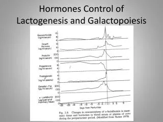
Hormone Priming on Lactogenesis From:Sheffield, l.G. and C.W. Welsch, J. Dairy Sci, 71:75-83, 1988.
Growth Hormone Levels in Cattle Selected for Higher Milk Production vs. Control Cattle
Oxytocin • Oxytocin is a 9 amino acid long peptide. The amino acid structure of oxytocin is: • Cys-Tyr-Ile-Gln-Asn-Cys-Pro-Leu-Gly • It has a molecular mass of 1007 daltons. Oxytocin has a disulfide bond between the two cysteines. Reduction of the disulfide bond inactivates oxytocin. One IU (international Unit) is approximately 2 micrograms of pure peptide.
Oxytocin Synthesis • Oxytocin is synthized in the hypothalamus in specific nuclei, the paraventricular nucleus and the supraoptic nucleus in the hypothalamus. [A cluster of nerve cells in the brain is often called a nucleus. This is different from the nucleus of a single cell.] Neurons in these hypothalamic nuclei synthesize the oxytocin precursor and package it into vesicles. Oxytocin is initially synthesized as a large molecular weight precursor which also consists of the oxytocin-carrier peptide neurophysin. The precursor is proteolytically cleaved in the neuron in the oxytocin-containing vesicle to yield oxytocin bound to neurophysin. The oxytocin-neurophysin complex is the intracellular storage form of oxytocin. • The oxytocin-containing vesicles are transported from the cell body (which is in the hypothalamus), down the axons to the neuron endings in the posterior pituitary. This is called the hypothalamo-neurohypophysial tract. The oxytocin-neurophysin complex is stored in neurosecretory granules called herring bodies in the axon ending.
Alveolus Stained to Show Myoepithelium Myoepithelial Cell
Oxytocin Release and Half-Life • It is estimated that the bovine pituitary has about 800 micrograms of oxytocin. This is about 40X what is in the blood under resting conditions. Only about 1/3 of pituitary oxytocin is released at a milking. • Oxytocin receptors on myoepithelial cells can respond to very low levels of oxytocin. • Oxytocin has a short half-life in the blood = 0.55 to 3.6 min. This means that the removal of milk by machine or by nursing must be closely timed with stimulation of the teats.
Factors Modifying Milk Letdown • Autonomic nervous system • Stress gives epinephrine release • Inhibits oxytocin release • Inhibits myoepithelial cell contraction • Inhibits blood flow to udder • Conditioned reflex • Letdown in response to sights, sounds associated with milking
Adrenal Medulla Posterior Pituitary Udder Vasculature Ihnibits oxytocin release Inhibits Blood Flow Epinephrine Myoepithelium Inhibits Contraction Medulla Adrenal
Residual Milk • Left in udder after normal milking • About 10% of milk • Can remove with oxytocin
Removing Residual Milk • Oxytocin injections • Expensive • Not approved use • Machine stripping • High incidence of liner slips • Increases mastitis risk • Udder massage • Second oxytocin release.