Download
1 / 26
290 likes | 614 Vues
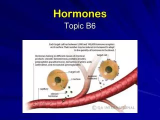
Hormones of pregnancy. Pregnancy. Preparation of uterus Steroid hormones Fertilization Coitus Gamete transfer Capacitation of sperms Fusion of gamates. Embryonic development Preimplantation Implantation Placentation Differentiation of cells Organogenesis.

E N D
Pregnancy • Preparation of uterus • Steroid hormones • Fertilization • Coitus • Gamete transfer • Capacitation of sperms • Fusion of gamates
Embryonic development • Preimplantation • Implantation • Placentation • Differentiation of cells • Organogenesis
Must alter cyclic changes in the ovarian steroid hormones • Progesterone • High • Must maintain the CL • Most species • Some can maintain pregnancy without CL after certain stage (placental progesterone)
Luteolysis • Destruction of the CL • Reinitiation of reproductive cycle • Two types • Active • Passive • Active luteolysis • Production of luteolytic agent (PGF2a) • Uterus • Passive luteolysis • Loss of luteotropic agents
Active luteolysis Ovarian artery • Communication from uterus to ovary • Approximately 4 days before estrus, the uterus begins to produce PGF2a, • PGF2adiffuses into the bloodstream feeding the ovary bearing the CL (ovarian artery). Uterine vein PGF2a Large black arrows indicate direction of PGF2a flow
Progesterone • From uterus to ovary • Interaction between PGF2a and its receptor • Large luteal cells • Decreased production of progesterone • Death of the luteal cells • Elevated intracellular Ca level • Constriction of blood vessels • Release of oxytocin. Oxytocin PGF2a PGF2a
Progesterone • From ovary to uterus (and back to the ovary) • Oxytocin • Reaches the uterus and stimulates production of more PGF2a • Increasing amount of estradiol from the large follicle • Increased production of PGF2a by uterus through increased sensitivity to oxytocin Oxytocin PGF2a PGF2a
Progesterone • From ovary to uterus (and back to the ovary) • Positive feedback loop • Uterine production of PGF2a • Production of oxytocin by the CL • Ultimately leads to corpus luteum regression • Reinitiation of reproductive cycle Oxytocin PGF2a PGF2a
Local regulation of reproductive cycle Progesterone • Progesterone production by CL • Begins to decline. • Initiated by increased production of PGF2a • Increased production of PGF2a • Ablated when pregnancy has been initiated, resulting in continued Progesterone production by the CL and pregnancy maintenance Pregnancy PGF2a
Maternal recognition of pregnancy • Two types • Anti-luteolytic • Diversion of PGF2a secretion • Inhibition of PGF2a secretion • Luteotropic • Maintenance of the CL by providing necessary hormone • Gonadotropin
Early embryonic development Uterotubal Junction Ampullary- isthmic Junction Isthmus Ampulla • Zygote • Begins to divide as it moves through the oviduct towards the uterus • Numbers of cells increase after each division • The size of the embryo does not (cell size decreases by approximately 20 % after each division)
Early embryonic development 8-cell embryos 2-cell embryo • Cells of the embryo remain within the zona pellucida as they divide • The size of the nucleus increases • All chromosomes remain intact • In cows, the embryo divides three to four times (approximately one division a day) while in the oviduct • Usually at the 16-cell or morula stage when it reaches the uterus
Early embryonic development • Morula stage • All the cells of the embryo are in a tightly packed clump • Cells on the inside of the clump • Different from those on the outside • Cells inside begin to further pack themselves together and form a mass of cells called the inner cell mass (ICM), located at one end of the embryo Morula-stage embryo Blastocyst-stage embryo ICM Blastcoele
Early embryonic development • The ICM • Develops into the fetus • The outer layer of cells lining the zona pellucida • Trophoblast • Placenta • Formation of a fluid-filled cavity • Blastcoele • Blastocyst Morula-stage embryo Blastocyst-stage embryo ICM Blastcoele
Early embryonic development • Cells in the ICM and trophoblast • Continue to divide • Blastacoele continues to accumulate fluid • Hatching • Floats freely until it attaches itself within lumen of the uterus Hatched blastocyst Zona
Attachment and establishment of pregnancy Embryo ICM ICM Placenta • After hatching • Rapid growth and development phase. • In cows, the blastocyst begins to rapidly elongate around 13 days after estrus, transforming from an ~3 mm spherical blastocyst into a long, thread-like form (around 25 cm in length) in 3 to 4 days • The elongation of the bovine embryo • Due to rapid proliferation of trophoblast cells • Cells in the ICM divide slowly during elongation
Attachment and establishment of pregnancy Inner cell mass Uterine endometrium Trophoblast layer • Cattle and sheep • Attachment of trophoblast to the uterine wall • Superficial with some fusion between uterus and trophoblast cells
PGF PGF PGF PGF Implantation and establishment of pregnancy Non-Pregnant • Conceptus (embryo plus placental tissue) • Produces interferon-tau (IFN-t) as it elongates • Prevents production of PGF2a by endometrium of the uterus Endometrium PGF Uterine vein PGF PGF PGF Pregnant Conceptus IFN-t IFN-t IFN-t IFN-t Endometrium Uterine vein
Diversion of PGF2a secretion • Pigs • Non-pregnant • Endocrine factor • Conceptus • Divert secretion(exocrine) • Estradiol • Increased production during 11-12 days post coitus • Conceptus
Diversion of PGF2a secretion • Local factor rather than systemic factor • Conceptus must be present in both uterine horns
Secretion of luteotropic substances • Species with passive luteolysis • Primates • Secretion of glycoprotein hormone • Syncytiotropoblast • Human chorionic gonadotropin (hCG) • Basis of pregnancy test • Secretion begins around 10 days after ovulation
hCG • Luteotropic hormone • LH-like activity • Binds to LH receptors in the CL • Maintenance of progesterone production • Increased lifespan during early stage of pregnancy • Production • Peaks around 9 to 14 weeks of pregnancy • CL loses its function during this time • Switch in steroidogenesis (placenta) • Declines gradually thereafter
Neuroendocrine system • Rodents and rabbits • Coitus as stimulus • Physical contact • Physical stimulation of reproductive tract (cervix) • Release of prolactin by the anterior pituitary gland
Neuroendocrine system • Prolactin • Luteotropic hormone • Switch to placental hormones • Placental lactogen • CL • Eventually dies • Steroid production by placenta
Horses • Recognition of pregnancy • Movement of embryo within the uterus • 12-14 times a day during day 12-14 of pregnancy • Eventual lock-down of the embryo • Production of glycoprotein • eCG • Cause luteinization of the large follicle • Formation of secondary CL • FSH-like activity in other mammals • Loss of both CLs • Placental progestigens