Hormones
Hormones. Chapter 12. Endocrine System Function. Major control and communication system Controls activities that require long duration digestion and energy metabolism osmoregulation, water balance, ion balance and excretion growth and development reproduction. Endocrine Systems.

Hormones
E N D
Presentation Transcript
Hormones Chapter 12
Endocrine System Function • Major control and communication system • Controls activities that require long duration • digestion and energy metabolism • osmoregulation, water balance, ion balance and excretion • growth and development • reproduction
Endocrine Systems • Invertebrates • Most hormones arise from neurosecretory cells in CNS • Hormones typically released directly to target tissues • Principally involved in regeneration, growth, development, and reproduction • Little homeostatic function
Endocrine Systems • Vertebrates • Much greater prevalence of non-neural endocrine glands • More complex control pathways • One hormone stimulates the release of another. • Greater involvement in homeostasis
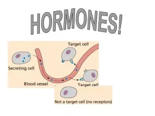
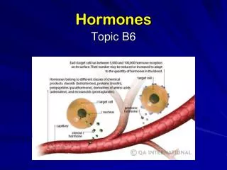
Hormones • Chemical signals broadcast to other cells • Endocrine • signals circulated throughout the body • Paracrine • signals broadcast locally within tissues • Autocrine • Signals act directly on the cell that releases it
Hormone Classes • Amines • hormones derived from tyrosine and tryptophan • adrenal medulla hormones, thyroid hormones, pineal gland hormones • Peptide Hormones • made from polypeptide chains • most hormones (insulin, FSH) • Steroids • derivatives of cholesterol • adrenal cortex hormones, gonadal hormones Figs. 14.1-14.3
Mechanism of Action:Steroids & Thyroid Hormones • nonpolar • pass directly through the cell membrane • bind to protein receptor in cytoplasm or in nucleus • protein binds to gene on DNA in the nucleus • stimulates expression of that gene (protein production) Fig 2.23d
Mechanism of Action: Peptides and Most Amines • Polar • cannot pass through hydrophobic lipid bilayer • bind to receptor proteins on cell surface • activation of membrane-bound enzymes • production of a second messenger inside the cell • e.g. cAMP, DAG-IP3, • 2nd messenger activates or deactivates various enzymes Fig 2.26
Hormonal Regulatory Mechanisms • Regulating hormone levels • e.g. Negative feedback • Change causes change in opposite direction • e.g. thyroxine/TSH • Regulating tissue response • e.g. down regulation • Decrease # of receptors on target cell with chronically elevated hormone levels Ex. Fig 14.8
Vertebrate EndocrinologyHypothalamus-Pituitary Axis • Hypothalamus (brain region) • controls release of pituitary hormones • Neural control of endocrine function • Pituitary gland • Two distinctive lobes (posterior and anterior) • Linked to hypothalamus by infidiubulum Figs. 10.8, 14.6
Posterior Pituitary (Neurohypophysis) • Composed of neurosecretory cells • Hormones released when neurons undergo action potentials Fig 14.6a
Posterior Pituitary Hormones • ADH (Anti-Diuretic Hormone) • Arginine vasopressin (mammals) or arginine vasotocin (other verts) • increases reabsorption of H2O by kidneys • induces vasoconstriction in arterioles - BP • Sexual behavior (amphibians) and oviposition (reptiles and birds) • Skin permeability (amphibians) • Oxytocin • Uterine contraction during childbirth • milk letdown during breast feeding • male function unclear ( occurs in both sexes during sexual arousal)
Anterior Pituitary (Adenohypophysis) • Composed of epithelial cells • Different cell types secrete various peptide hormones • Secretion controlled by hormonal release from hypothalamus into hypothalamal-hypophyseal portal system Fig 14.6b
Anterior Pituitary Hormones • TSH (Thyroid Stimulating Hormone, Thyrotropin) • Stimulates thyroid gland • Release thyroid hormones • Stimulates thyroid growth • ACTH (Adrenocorticotropin) • Stimulates adrenal cortex to release glucocorticoids Fig 14.6b
Anterior Pituitary Hormones • PRL (Prolactin) • Breast development and milk production during pregnancy • Development and maintenance of corpus lutea (non-primate mammals) • Crop milk secretion in pigeons, brood patch development in birds • Controls sensitivity of testes to LH • Enhances uptake / inhibits secretion of ions in fish and amphibians • Lots of other modulatory functions Fig 14.6b
Anterior Pituitary Hormones • MSH (Melanocyte Stimulating Hormone) • Integument pigmentation • GH (Growth Hormone, Somatotropin) • Stimulates growth, protein synthesis, fat breakdown and blood glucose levels • Functions indirectly through somatomedins (e.g., insulin-like growth factors) Fig 14.6b
Anterior Pituitary Hormones • FSH (Follicle Stimulating Hormone, Follitropin) • regulates female sex hormones, egg development • Stimulates Sertoli cells to release local mediators that induce spermatozoa development • LH (Luteinizing Hormone, Lutropin) • ovulation, regulation of female sex hormones • induces corpus luteum formation after ovulation • Induces secretion of androgens by Leydig cells of testes Fig 14.6b
“Adrenal” Glands • Releases hormones in response to stress • glucocorticoids (e.g. cortisol) • Elevate blood glucose • Anti-inflammatory and Immunosuppression • mineralocorticoids (e.g. aldosterone) • Na+/K+ balance, blood pressure regulation • androgens (e.g., DHEA) • sexual characteristics • epinephrine (“flight vs. fight”) • ↑ blood glucose, lipolysis • ↑ thermogenesis (shivering and non-) • ↑ cardiovascular / respiratory activity Figs 14.5, 14.7
Thyroid Gland (Tetrapods) • Thyroid hormones (TH) • Thyroxine (T4) and triiodothyronine (T3) • Increase metabolic rate and body heat production (endotherms) • Metamorphosis in amphibians • Growth and development
Pancreas • Endocrine cells located in Islets of Langerhans • Contain two cell types • cells - secrete glucagon • cells - secrete insulin • Important in regulating glucose levels of the blood
Insulin • Induces glucose uptake and utilization by cells (esp. muscle and liver) • Lowers blood glucose levels • promotes removal of glucose from blood • Promotes formation of glycogen • polymer of glucose for storage • Promotes conversion of glucose into fat in adipose tissue • Stimulates amino acid uptake by cells and protein formation
Glucagon • Increase in blood glucose: • Activates liver enzymes to convert glycogen into glucose • Stimulates breakdown of stored fat and release of fatty acids into blood • used as secondary energy source
Gonads (Testes and Ovaries) • Produce steroid hormones • androgens (e.g., testosterone) • sperm development • reproductive tract maturation • secondary sex characteristics • sexual behavior (M and F) • estrogens and progesterone • oocytic development • reproductive tract development • secondary sex characteristics Figs 15.5c, 15.9b
Comparative Endocrinology:Insect Molting / Metamorphosis • Development patterns in insects: • Hemimetabolous • go through nymph stages (instars) and slowly transform into adults • Homometabolous • Go through prolonged larval stage, then develop into pupa, then adult • Development in both occurs through similar endocrine control Fig 14.6
Key Hormones • Prothoracicotropic Hormone (PTTH) • Peptide hormone secreted by neurosecretory cells brain • Stimulates release of ecdysone • Ecdysone • steroid hormone produced by prothoracic gland • Stimulates molting and development • Juvenile hormone (JH) • Terpenoid hormone produced by corpus allatum • Stimulates retention of juvenile characters Figs 14.18-14.19
Hormone Function: Molting • Growth of juvenile stimulates PTTH secretion • Stimulates ecdysis (molting) • Cuticle detaches from epithelium • Muscular contractions pull insect away from cuticle • New cuticle forms Fig 14.19
Hormone Function: Metamorphosis • Levels of JH initially high • Retains juvenile characters • Levels begin to fall as larva grows • When JH falls to below a certain level → pupation • Continue to fall to during pupation • At minimum JH production, adult form develops • JH levels rise in adult • stimulate reproductive development
Additional Important Hormones • Eclosion Hormone (EH) • stimulates inka cells to secrete PETH and ETH • Pre-ecdysis Triggering Hormone (PETH) • Coordinates muscle contractions that pull epidermis away from old cuticle • Ecdysis Triggering Hormone (ETH) • Cooordinates contractions that allow final escape from the old cuticle • Bursicon • tanning/hardening of new cuticle after molting Fig 14.19