Nucleic Acids (Chapter 26)
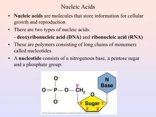
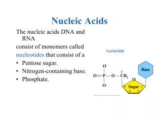
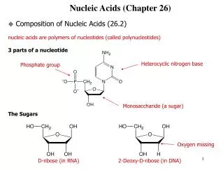
Nucleic Acids (Chapter 26). Composition of Nucleic Acids (26.2) nucleic acids are polymers of nucleotides (called polynucleotides) 3 parts of a nucleotide The Sugars. Heterocyclic nitrogen base. Phosphate group. Monosaccharide (a sugar). Oxygen missing. D-ribose (in RNA).

Nucleic Acids (Chapter 26)
E N D
Presentation Transcript
Nucleic Acids (Chapter 26) Composition of Nucleic Acids (26.2) nucleic acids are polymers of nucleotides (called polynucleotides) 3 parts of a nucleotide The Sugars Heterocyclic nitrogen base Phosphate group Monosaccharide (a sugar) Oxygen missing D-ribose (in RNA) 2-Deoxy-D-ribose (in DNA)
The Bases Adenine DNA, RNA Guanine DNA, RNA Cytosine DNA, RNA Thymine DNA Uracil RNA 4 3 5 2 6 1 Sugar connects here pyrimidines purines Sugar + Base = Nucleoside 6 7 Dehydration synthesis - loss of water 5 Change names: Adenosine Guanosine Thymidine Cytidine Uridine 1 8 2 4 9 3 5’ + 4’ 1’ 2’ 3’ Ribose Adenine Adenosine (a nucleoside)
Sugar + Base + Phosphate = Nucleotide Building blocks of nucleic acids - monomers of DNA and RNA named by adding 5’ monophosphate at the end of the nucleoside name. Adenosine 5’ monophosphate, guanosine 5’ monophosphate, etc Adenosine (a nucleoside) Adenosine 5’-monophosphate (a nucleotide) Ribonucleotide vs. deoxyribonucleotide Ribonucleotide - contains ribose used for RNA deoxyribonucleotide - contains deoxyribose used for DNA
O O N N - - O O P P O O C C H H O 2 2 O O N O O - - - O P O C H 2 O O O H H O - O N O P O C H 2 O O - O H Structure of Nucleic Acid Chains (26.3) nucleotides are linked together through phosphodiester linkages bond between 3’ OH group of nucleotide and the phosphate of the next Note: pg. 745 fig is wrong - the P is not dbl bond to the 3’ O 5’ end 5’ 5’ 3’ + 5’ Phosphodiester link 3’ 3’ 3’ end A dinucleotide
The phosphates and sugars form the DNA backbone DNA sequences differ based on the order of the bases like protein sequence differs based on the order of the side chains A DNA sequence is read from the 5’ to the 3’ end
Draw the full structure of the trinucleotide C-A-T and label the 5’ and 3’ ends. Draw a box around each nucleotide.
Base Pairing in DNA (25.4) Chargaff’s Rules - %A = %T, %G = %C DNA consists of 2 polynucleotide strands non-covalently interacting Strands wind around each other in a helical, screw-like fashion - called the double helix Sugar-phosphate backbone is on outside, bases are on inside resembles a spiral staircase The hydrophobic effect holds the 2 strands of DNA together - NOT H-BONDS H-bonds and ion-dipole interactions with cations and solvent add to stability
Watson-Crick Base-Pairing H-bonds are responsible for directing replication and transcription base pairing is described as complementary. If T is on one strand, A is on the opposite. 2 H-bonds 3 H-bonds
Structure of duplex DNA 2 Chains run antiparallel 3 types of duplex DNA 1) B-DNA - right handed helix - most studied/common form in body 10 base pairs per turn 2) A-DNA - right handed helix - found in low salt conditions 11 base pairs per turn 3) Z-DNA - left-handed helix - found in nature - function unknown occurs in high G/C areas B-DNA wide major groove in B-DNA and narrow minor groove many proteins such as DNA polymerase, RNA polymerase bind in the major groove major minor
Organization of DNA Total number of base pairs in a human cell is 3 billion Chromosome - molecule of DNA In humans - 46 chromosomes - 23 pairs of chromosomes horse - 64 chromosomes (32 pairs) cat has 38 (19 pairs) mosquito has 6 (3 pairs) If you straighten out all the DNA in a cell and line up end to end, there is about 2 meters of DNA in each cell Gene - each chromosome is made up of thousands of genes - estimated there are ~40,000 genes - codes for a protein genetic code (26.9) - sequence of the bases specifies the sequence of amino acids in a protein codon - base triplet that codes for an amino acid insert codon table
Replication of DNA (26.6) DNA double helix partially unwinds Bases are added based on the opposing strand as nucleoside triphosphates DNA polymerase is the enzyme that copies DNA - it catalyzes the formation of the phosphodiester bond between the 3’ OH and the 5’ phosphate on an incoming nucleoside triphosphate New DNA is synthesized from the 5’ to the 3’ direction New strand growing OH attacks P Bond breaks releasing a lot of energy
Replication is semi-conservative Each strand in the parent DNA acts as a template for the synthesis of a new DNA strand. End up with 2 new identical DNA molecules with one parent strand per new molecule and one new strand per new molecule DNA always made in 5’ to 3’ direction and strands are always antiparellel
Transcription of DNA (26.8) Transcription - synthesis of messenger RNA (mRNA) using DNA as a template occurs in the nucleus, 1st step in protein synthesis DNA section to be transcribed is unwound only 1 strand of DNA is used as a template (template strand) the mRNA produced is complementary to the template strand but identical to the non-template DNA strand (called the informational strand) (except U for T and sugar) mRNA is produced from the 5’ to the 3’ end like in replication purpose of mRNA is to carry genetic information from DNA to cytosol Example: Write the DNA complement (template strand) and the mRNA strand DNA strand (informational strand) 5’ ATG CCA GTA GGC CAC TTG TCA 3’ DNA strand (template strand) 3’ TAC GGT CAT CCG GTG AAC AGT 5’ mRNA 5’ AUG CCA GUA GGC CAC UUG UCA 3’ mRNA made is a complete copy from start to stop codon not everything copied is used to make protein Exons are in pink - expressed part of gene intervening parts that do not code for protein This is the mRNA that is used to make protein
Translation of mRNA (26.10) Translation - synthesis of protein based on mRNA sequence mRNA leaves nucleus - translation occurs in the cytoplasm on ribosomes ribosome - scaffolding for translation - holds the mRNA, tRNA, and enzymes in place composed of rRNA and protein t-RNA - each codon has a t-RNA with the corresponding amino acid covalently attached anticodon is at base of t-RNA molecule and is complementary to codon on mRNA - the is responsible for bringing the correct amino acid to the ribosome to add to the protein chain. F in yellow aa binds here anticodon
Genetic Mutations Point Mutations- a single base change Normal DNA ATG-GAC-TTC Mutant DNA ATG-CAC-TTCClassified as: transitions- flipping the base pair to complement DNA: G to C or A to T. (purine purine or pyrimidine pyrimidine) transversions- switching base pairs: G/C to A/T. (purine pyrimidine or vice versa) Is the above mutation a transition or transversion? Point mutations may or may not affect transcription: Missense mutations- a change that specifies a different AA. Ex. GUU GCU resulting in Val Ala Base substitution leads to AA substitution. May result in no phenotype, mild, or very serious consequences. Refer to pg. 757 table mRNA codon assignments of base triplets. Normal DNA A T G - G G C - T T C RNA A U G – G G C – U U C Amino acid _Met_ - _Gly_ - _Phe_
Nonsense mutations- a change that produces a stop codon resulting in a prematurely • shortened protein. • ex. CGA UGA gives Arg stop • The effects are variable depending upon how much truncated protein is present and • required for its function. • Normal DNA T T G - A A C - T A C • RNA A A C - U U G - A U G • Amino acid _Asn_ - _Leu_ - _Met_ • Normal DNA T T G - A T C - T A C • RNA A A C - U A G - A U G • Amino acid _Asn_ - _stop_ - _Met_
Silent mutations causes a change that specifies the same AA. Ex. GUU GUC results in Val Val How is that possible? (a) Often true in 3rd position of a codon, especially transitions. The change doesn’t Modify the codon to produce a different AA ( single- nucleotide polymorphism (SNP) specifies The same AA. (b) May terminate protein synthesis by introducing a stop codon. the genetic code is degenerate result Normal DNA T T G - A T C - T A C RNA A A C - U A G - A T G Amino acid _Asn_ - _stop_ - _Met__ Normal DNA T T G - A T T - T A C RNA A A C - U A A - A T G Amino acid _Asn_ - _stop_ - _Met_
Frameshift mutations- the number of the inserted or deleted bases Not in a multiple of 3, so that all triplets following the mutation are Read differently. (a) this mutation changes all the AA downstream & very likely to create a nonfunctional product since it may differ greatly from normal protein. (b) reading frames other than the correct one often contain stop codons which will truncate the mutant protein prematurely. Deletion Mutation * loss of one or more bases Normal DNA A T G - G A C - T T C Mutant DNA ATG – GCT- TC Insertion Mutation * addition of one or more bases Normal DNA A T G - G A C - T T C Mutant DNA ATT - GGA – CTT- C
Causes of Mutations Can be the result of different events: 1) Spontaneous- occurs as a result of natural processes in the cell. - naturally occurring mutagens in the environment - DNA replication errors 2) Mutagens- external agent that can cause a mutation (induced) - error in base sequence that is carried along in DNA replication. Carcinogens – cancer causing agents/ chemicals in the environment. ( contain polycyclic aromatic hydrocarbons and amines) Examples: lung cancer from chemicals in cigarettes
Sunbathing damages DNA UV radiation does numerous things to DNA 1) Causes breaks in sugar phosphate backbone - DNA cannot replicate/transcribe - results in cell death 2) Causes “photodimers” - thymine dimers are most common - occurs btw adj T on same strand
Thymine dimers “kink” DNA Undamaged DNA UV damaged DNA thymine dimer pulls thymines closer together causing the DNA kink The kink causes: 1) replication/transcription to terminate at the point of the dimer or even worse, 2) DNA polymerase iota will replicate through but misinserts T or G opposite one of the thymines in the dimer instead of A and only 1 base is inserted opposite it.
Cigarette smoke damages DNA Benzo[a]pyrene is the main cancer causing agent in cigarette smoke Benzo[a]pyrene enters body and is converted to BPDE which covalently attaches to DNA This is your DNA This is your DNA on Cigarettes
How does BPDE cause mutations? p53 - tumor suppressor protein - scans DNA for damage during cell division - if damage is spotten, p53 stops cell division and activates DNA repair systems - if damage is too severe to be fixed, p53 triggers apoptosis (programmed cell death) - over 50% of all cancers have a disabled p53 - over 80% of all lung cancers have a disabled p53 BDPE is a potent mutagen of p53 by causing G:C to T:A transversions