Download
1 / 118
2.55k likes | 7.81k Vues
What are Intravenous fluids ?. Are large volume injections intended to be administered by intravenous infusion. •Included in the group of sterile products referred to as Large Volume Parenterals (LVPs).

E N D
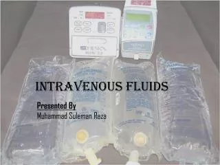
What are Intravenous fluids? • Are large volume injections intended to be administered by intravenous infusion. • •Included in the group of sterile products referred to as Large Volume Parenterals (LVPs). • Consists of single-dose injections having a volume of 100mL or more and containing no added substances.
Are sterile solutions of simple chemicals such as sugar, amino acids, or electrolytes-materials that easily can be carried by the circulatory system and assimilated. • Prepared with Water for Injection, USP • Pyrogen-free solutions • A solution (usually a balanced electrolyte solution) administered directly into the venous circulation.
What are the different types of intravenous fluids? • IV fluids can be classified into: • •Crystalloid Solutions • •Colloidal Solutions
Crystalloid Solutions • Crystalloid Solutions: • Contain small molecules that pass freely through cell membranes and vascular system walls. • Are primary fluids used for IV therapy. • These solutions are useful as fluid expanders and are stored at room temperature. • Useful source for electrolytes and a temporary source of fluid volume
Saline Solutions • Saline solution is a solution of sodium chloride, or salt, in sterile water. • A. 0.9% Normal Saline: • contains 9g/L sodium chloride • Has osmolarity of 308 mOsmol/L (calc). • It contains 154mEq/L sodium and 154mEq/L chloride. • Is a solution commonly used for medical purposes such as intravenous therapy for severe dehydration. • It is also used as a rinse for contact lenses, and is used in wound care for irrigating, cleansing and hydrating wounds. • Has green label.
B. 0.45% Normal Saline Solution: • Hypotonic Saline. • Contains 4.5g/L Sodium Chloride. • Has osmolarity of 154 mOsmol/L (calc). • It contains 77mEq/L sodium and77mEq/L chloride. • C. 1.8,3.0,7.0,7.5 and10% Saline Solution: • Hypertonic Saline
Different volumes of IV bags are used in the pre-hospital environment.
Expiration date Fluid type 250 ml
Dextrose solutions • Dextrose Solutions- used to supply water and calories to the body. • It is also used as a mixing solution (diluent) for other IV medications. • A. 5% Dextrose (D5W): is a parenteral fluid and nutrient replenisher. • Each100mL of 5% Dextrose Injection, USP, contains dextrose, hydrous 5g in water for injection. The caloric value is 170 kcal/L. • The osmolarity is 252 mOsmol/L (calc.), which is slightly hypotonic. • Has red label • B. 5% Dextrose in 0.9% Saline (D5NS): is a sterile, nonpyrogenic solution for fluid and electrolyte replenishment and caloric supply in single dose containers for intravenous administration. • Has yellow label.
C. a 5% dextrose in0.45%Saline(D51/2NS) • D. Dextrose 5% in Lactated Ringer’s (D5LR) • has purple/pink label.
Lactated Ringer’s Solution • Lactated Ringer’s Solution-is an intravenous (IV) solution used to supply water and electrolytes (e.g., calcium, potassium, sodium, chloride), either with or without calories (dextrose), to the body. • It is also used as a mixing solution (diluent) for other IV medications. • Has blue label.
Colloid Solutions • It contains molecules that are frequently very complex and much larger than those in the crystalloid solutions. • It is needed when a solution is required to remain in the vascular system. • It is generally require refrigeration and can be stored for a limited period. • Whole human blood U.S.P. and Heta starch are examples of colloid solutions. • .
What are the different components of an IV fluid? • Water for patients with dehydration • Amino Acids-for tissue growth and repair, replacing body cells, healing wounds, and synthesizing vitamins and enzymes. • Vitamins(A, D, E, K, B&C)-for the rest or active and replacement therapies. • pH-for the acidity and alkalinity of a solution
Electrolytes-major additives for replacement and restorative therapies. • -any compound that, in solution or in molten form, conducts electricity and is decomposed (electrolyzed) by it. It is an ionizable substance in solution • -is any substance that contains free ions that behaves as an electrically conductive medium (conducts electricity).
Electrolytes • 1- Sodium • Functions : Regulation for water regulation. It helps with electrical signals in the body, allowing muscles to fire and the brain to work. • Sources: Sodium Acetate, Sodium Phosphate. 2- Potassium: • Function: Regulation of acid-base balance •It is essential in the generation of the electrical impulses in the body that allow muscles and the brain to function • Sources: Potassium Chloride, Potassium Phosphate • 3- Magnesium: • Function: is involved with a variety of metabolic activities in the body, including relaxation of the smooth muscles that surround the bronchial tubes in the lung, skeletal muscle contraction, and excitation of neurons in the brain. •It acts as a cofactor in many of the body's enzyme activities. • Sources: Magnesium Sulfate, Magnesium Phosphate
4- Calcium: • Function: Used in building and maintaining bones and teeth. Aids in blood clotting, nerve function, and muscle contraction. Maintains normal levels of blood pressure and stomach acids. • Sources: Calcium Gluconate, Calcium Chloride
5- Phosphate: • Function: • It helps form strong bones and teeth in the human body. • It helps filter waste from the kidneys and plays a vital role in the production and storage of energy in the body. • It is responsible for maintaining the balance of other nutrients since it combines with other minerals to form phosphate salts or compounds • Sources: Phosphate salts of sodium and potasium.
6- Chloride: • Function: • It travels primarily with sodium and water and helps generate the osmotic pressure of body fluids. • It is an important constituent of stomach hydrochloric acid (HCl), the key digestive acid. • It is also needed to maintain the body's acid-base balance . • It may also be helpful in allowing the liver to clear waste products. Sources: Chloride salt of cations
Nutrient Solutions: • carbohydrates(dextrose, glucose or fructose)-water • Example: • D5W (5% dextrose in water) • 5% dextrose in 0.45% NaCl (dextrose in half-strength saline) • Electrolyte Solutions:-cations and anions • Example: • NSS(0.9% NaCl solution) • Ringer’s Solution (Na, Cl, K and Ca) • Lactated Ringer’s Solution( Na,Cl,K,CaandLactate)
Alkalanising Solutions-for metabolic acidosis • Acidifying Solutions -for metabolic alkalosis • Blood Volume Expanders • Example: • Dextran • Plasma • Human serum albumin
Uses of IV Fluids • Intravenous fluids commonly are used with the following conditions: • 1- Correction of disturbances in electrolyte balance (Na,K,Ca,PO4,Mg imbalance). • 2- Correction of disturbances in boy fluids (volume expander ,blood loss). • 2- Means of providing basic nutrition (provide patients with difficulty in taking food and fluids orally). • 3- Basis for the practice of providing Parenteral Nutrition. • 4- Vehicles for other drug substances (mixed with fluids for medication needed in the body)
Define: • Hypertonic- is a solution having a larger concentration of a substance than is found within the cells themselves.-it causes the cell to shrink , or crenate. • Hypotonic- it contains a lesser concentration of impermeable solutes on the external side of the membrane.-it causes the cell to swell • Isotonic- a solution which has the same concentration of dissolved substances as the blood cells do.
What are the different types of IV administration sets and equipments? • IV Infusion can be administered either by: • 1) Gravity alone Example: • Gravity Infusion set. • 2)With the use of an electronic infusion device Example: • Infusion pumps • Volumetric pumps
1) IV infusion administered by gravity: • Gravity Infusion Set: • The height of the IV solution is of greater importance than the tubing. Most basic types of IV tubes/tubing can be used in this type of set. The higher the solution, the faster the solution infuses. Preferred elevation of the solution from the site of infusion: 18 to 24 inches (45 to 60 centimeters).
2) IV fluids administered with the use of an electronic infusion device: • Infusion Pump: • Pressure is used in order to infuse solutions Requires special tubing that contains a device such as cassette to create a sufficient pressure to push fluid into the vein. • Advantage: Programmed to deliver a preset volume per hour. • Disadvantage: If catheter or needle within vein becomes misplaced, the pump will still continue on infusing.
IV infusion administered with the use of an electronic infusion device: • Volumetric Pump: • Do not depend upon gravity to force the fluid into the vein. • All volumetric pumps generally involve the nurse entering the infusion rate in mL/hr. • The volumetric pump then automatically maintains that rate. • Volumetric pumps should still be checked regularly to ensure that they are infusing the medication correctly. • Infiltration is possible when using a volumetric pump because it forces the fluid into the vein, even when it encounters resistance.
IV Administration Equipments • Basic IV Set up consists of the following important parts/equipment: • a) Drip chamber b) Roller clamp c) Slide clamp d) Injection port • Other IV equipment : • a) IV Tubing b) Hypodermic needle • c) Catheter needle d) Central IV Lines • e)Tunneled Lines/Broviac Line • f)Peripherally inserted central catheter
Basic IV Equipment • A) Drip Chamber Located just below the IV bag. Inside this chamber, we can see the fluid drip down from the bag into the IV tubing. • This is where we measure the speed of a manual IV set up; we look at this chamber and count the number of drops we see per minute. • B) Roller Clamp: is what we use to control the rate at which the IV fluid infuses. • All roller clamps on a set of IV tubing should be closed before we attach a bag of IV fluid to the top of the tubing; this ensures that no air gets into the tubing
C) Slide clamp: slide clamp is used when we want to completely stop the IV from flowing without having to adjust the roller clamp. D)Injection port: A place where medicine or fluids other than those in the current IV bag can be injected so that they will infuse into the patient's vein through the IV tubing.
How the Height of the IV Bag Affects the Infusion Rate • IV infusion works because gravity pushes the fluid down through the IV tubing into the patient's vein. • The higher the bag is hung, the greater the gravitational pressure on the IV fluid to go downward through the tubing; • if the IV bag is not hung high enough, there will not be enough pressure caused by gravity to force the fluid into the vein. • So, all IV bags must be hung above the patient's heart in order for there to be enough pressure for the IV fluid to infuse, and it is standard procedure to hang the IV bag at least 3 feet above an adult patient's heart to ensure there is enough pressure to keep the IV running at a constant rate
A canula is a hollow needle, or more often a length of flexible plastic tubing which has been inserted into the vein using a needle; the tubing has been taped to the patient's arm to prevent it coming out when the patient moves, and a sterile dressing has been placed over the punctured place in the skin where the canula has been inserted to prevent bacteria that commonly exist on the skin's surface from getting into the bloodstream.
peripheral line • A peripheral line is an IV that is attached to a peripheral vein, which is any vein not located in the torso. These types of IV are usually inserted into the arm or hand, although a leg or foot may be used. This is the most common type of IV. • A peripheral line may only be used for a short period of time, usually 3 days, because if it is used for longer periods of time, bacteria that are normally present on the skin can travel into the blood or the tissue surrounding the injection site and cause infection. • So, if a peripherial line is needed for more than 3 days, it is standard procedure to move the injection site to a new location every 3 days to prevent infection.
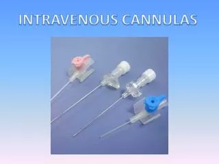
Other IV Equipments • A) IV Tubing Plastic (canula) conduit used to administer various fluids to patients through a needle inserted into one of the patient’s vein. • B)Hypodermic Needle Commonly used with a syringe to inject substances into the body or extract fluids from it. • C) Catheter Needle: • Used for the injection of fluids into the human body. • This device is widely used in hospitals.
D) Central IV Lines Have the capacity to deliver fluids that are considered to be irritating to peripheral veins. Also, medications introduced in this manner are quickly distributed throughout the body.
Central line • A central line is an IV that is attached to a vein in the chest. • Usually the canula is inserted through the chest wall or a neck vein, but it is also possible to insert the canula into a peripheral vein and then to move the tip of the canula slowly upward until it is in a central vein.
Central veins are much larger than peripheral veins, so when a central line is used and the canula is inserted through the chest or neck the tubing can be wider and so multiple smaller tubes can be inserted through the larger one to deliver several IV medications at once that are not allowed to mixed. • Also, a central line goes into a vein that carries blood directly to the heart, so medication given this way is distributed more quickly throughout the body. • Medicines that are particularly harsh or in a high concentration are also more likely to irritate a peripheral vein, such as chemotherapy drugs and some kinds of liquid nutrition, can be given in a central line when they are too irritating to be administered via a peripheral vein.
However, a central IV line is also more likely to cause bleeding and the risks of infection are much higher because the contents of the line go directly to the heart, so any bacteria that get into the line are quickly spread throughout the body; also, the risks of getting air in the line that could block veins or stop the heart are higher with a central line since it is wider and therefore allows for larger amounts of air to enter (the more air that enters the bloodstream, the greater the danger that a vein will be blocked or the heart will stop).
Continueous versus Intermittent IV Infusion • Sometimes an IV medication or fluid is given continuously, or all the time. But sometimes we may want to administer an IV fluid and/or medication to a patient only at specific times; this is called an intermittent IV infusion. A patient may receive only continuoeus IV fluids/medication, or only intermittent IV fluids/medication or a mixture of both. • A patient who is to receive a continuous IV has the IV setup connected to them all the time, but for a patient who should receive only intermittent IVs, we can't leave them permanently attached to an IV setup. What we do instead is insert a canula like the one in the picture before to the patient, which allows us to connect an IV only when the patient is actually receiving an infusion and to disconnect it in between doses.
This is a length of IV tubing with an injection port attached to one end; this special injection port is called an infusion port adapter, although it is also usually referred to as a heplock or a saline lock/port, because in an intermittent IV setup, the patient is not getting a constant flow of fluid through the canula, so it can become blocked by clotted blood and therefore must be flushed periodically in order to clear it out; heparin (in a concentration of 100 U/mL), a drug which prevents blood from clotting, and saline, or salt water, are the two fluids that are used for this flush, which involves an injection of approximately 1-2 mL of either of these fluids every 6 to 8 hours.