Guideline for gout management
370 likes | 1k Vues
Guideline for gout management. (Arthritis). 高雄長庚醫院風濕過敏免疫科. Introduction. the deposition of monosodium urate ( MSU ) crystals in the joints and soft tissues. Incidence: 0.1%. Introduction.

Guideline for gout management
E N D
Presentation Transcript
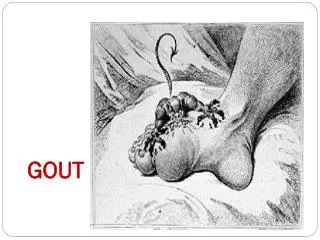
Guideline for gout management (Arthritis) 高雄長庚醫院風濕過敏免疫科
Introduction • the deposition of monosodium urate ( MSU ) crystals in the joints and soft tissues. • Incidence: 0.1%
De novo and salvage pathways in purine metabolism. Phosphoribosyl pyrophosphate amidotransferase (AMPRT) catalyzes the committed step of de novo purine nucleotide synthesis. Hypoxanthine phosphoribosyltransferase (HPRT) and adenine phosphoribosyltransferase (APRT) are responsible for recycling purine bases into nucleotides. 5-phosphoribosyl-1-pyrophosphate (PRPP) levels regulate all of these reactions. Uricase (UC) prevents the buildup of uric acid in mice, but not in humans. Other important enzymes in the salvage pathway are adenosine deaminase (ADA), purine nucleoside phosphorylase (PNP), guanase (GA), and xanthine oxidase (XO).
Clinical course 4 clinical phases if untreated: • asymptomatic hyperuricemia, • acute/recurrent gout, • intercritical gout, • chronic tophaceous gout
Asymptomatic Hyperuricemia • elevated urate levels without symptoms of gout, nephrolithiasis, or kidney stones. • Hyperuricemia is defined: >7 mg/dL (0.42mmol/L) in men and postmenopausal women >6 mg/dL (0..36mmol/L) in premenopausal women. • urate <7 mg/dL 0.1% annual incidence of gout urate >=9 mg/dL 4.9% annual incidence. • the clustering of glucose intolerance, central obesity, dyslipidemia, hypertension, and increased prothrombotic and antifihrinolytic factors in an individual.
Primary Idiopathic Familial juvenile gouty nephropathy Secondary Hypertension Hyperparathyroidism Myxoedema Down’s syndrome Increased level of organic level Lead nephropathy Sarcoidosis Bartter’s syndrome Beryllium poisoning Drug: diuretics, B-blocker, ACEI, salicylates (low dose), PEA, EMB, cyclosporin, nicotinic acid Chronic renal failure Volume depletion NDI Cause of hyperuricemia-- decreased renal excretion
Primary Idiopathic HPRT def. PPRT overactivity Ribose-5-phosphate overproduction AMP-deaminase def. Secondary Glycogen storage disease type II (G6PD), type III, V, VII Hereditary fructose intolerance Lymphoproliferative and myeloproliferative diseases ( leukemia, Hodgkin’s d’z, lymphosarcoma, myeloma, PV, Waldenstrom’s macroglobulinemia ) Cytotoxic drugs Carcinomatosis Gaucher’s disease Chronic hemolytic anemia Severe exfoliative psoriasis Cause of hyperuricemia-- increased uric acid production
Acute/Recurrent Gout • symptoms: sudden onset of severe pain, inflammation, limited range of motion, and warmth at the affected joint(s). • slight fever, leukocytosis, elevation of ESR, and elevation of CRP • 90% of first attacks are monoarticular with first metatarsophalangeal joint, known as podagra. • Left untreated, the symptoms are self-limiting but may take up to 21 week to subside.
Intercritical Gout • After recovery from an acute gout flare, the patient enters an asymptomatic phase of the disease. • This interval between gout flares: as intercritical or interval gout. • Later, recurrence of acute gout may become more frequent and polyarticular involvement.
Chronic Tophaceous Gout • Tophi are usually present after 10 to 20 years of inadequately treated chronic gout. • Visible tophi occur in 12% of patients after 5 years of gout and in 55% of patients after 20 years. • most common sites of tophaceous gout: olecranon bursae (elbow) and the joints of the hand and feet. Other sites: the helix of the ear, the Achilles tendons, and the knees.
Complication of gout • Joint: destruction • Soft tissue • nerve entrapment syndrome: CTS, tarsal tunnel syndromes • kidney: uric acid calculi(10-15%), chronic urate nephropathy, and acute uric acid nephropathy • Heart: ischemic heart disease
Criteria for clinical diagnosisAmerican Rheumatism Association sub-committe on classification criteria for gout 1977 • presence of characteristic urate crystals in the joint fluid • Tophus proved to contain urate crystals by negative polarized light microscopic study • If none of above, diagnosis is 6/12 clinical, radiographic, and laboratory criteria include: 1. more than one attack of acute arthritis 2. Maximum inflammation within 24 hours 3. Attack of monoaricular arthritis 4. Joint redness observed 5. first MTP joint painful or swollen 6. Unilateral attack involving first MTP 7. Unilateral attack involving tarsal joint 8. Suspected tophus 9. Hyperuricemia 10. Asymmetric swelling within a joint ( roentgenogram ) 11. Subcortical bone cysts without erosions ( roentgenogram ) 12. Negative synovial culture during attack of joint inflammation
Acute Infective arthritis Bursitis, cellulitis, tenosynovitis Other crystal arthropathy ( pseudogout, apatite or brushite arthritis or periarthritis ) Traumatic arthritis Hemoarthrosis RA with palindromic onset Reactive arthritis Spondarthritis with peripheral involvement Psoriatic arthritis Sarcoid arthritis Rheumatic fever Chronic Nodular rheumatoid arthritis Psoriatic arthritis Osteoarthritis with Heberden’s and Bouchard’s nodes Sarcoid arthritis xanthomatosis Differential diagnosis
History taking • Age of onset • Involving joints • Frequency of attack • Family hx • Previous treatment and other medication • Associated medical hx: 4H ( hypetension, hyperglycemia, hyperlipidemia, and hyperuricemia )
Events provoking acute gouty arthritis • Trauma • unusual physical exercise • Surgery • Severe systemic illness • Severe dieting • Dietary excess • Alcohol • Drugs ( diuretics, initiation of uricosuric or allopurinol therapy, initiation of B12 therapy in pernicious anemia, cytotoxic drug therapy )
Physical examination • Vital sign • Body weight and body height • General appearance: Cushingnoid … • Consciousness • HEENT • Chest ( CV ) • Abdomen • Extremity -- PE of joint: appearance, joint effusion, ROM -- location of tophi -- sign of neuropathy -- muscle power, DTR…
Diagnostic evaluation • CBC/DC • Glucose, Na/K, Ca/P, uric acid, AST/ALT/ALP, HDL-cholesterol electrophoresis • U/A, 24hr uric acid(U) • Synovial study • Special investigation -- EKG, CXR, joint radiography -- skeleto-muscular ultrasound examination
Long-term treatment • Indication: 1. Recurrent attacks 2. Evidence of tophi or chronic gouty arthritis 3. Associated renal disease 4. Patient is young with high serum UA and FH of renal or heart disease 5. Normal serum UA cannot be achieved by life-style modifications • Medication: 1. Allopurinol 2. Uricosuric agents: probenecid or sulfinpyrazone 3. benzbromarone
Indications for Antihyperuricemic Therapy in Gout • Frequent and disabling attacks of acute gouty arthritis • Clinical or radiographic signs of chronic gouty joint disease • The presence of tophaceous deposits in soft tissues or • subchondral bone • Gout with renal insufficiency • Recurrent nephrolithiasis • Serum urate levels persistently in excess of 13 mg/dL in men • or 10 mg/dL in women • Urinary uric acid excretion exceeding 1100 mg/day • Impending cytotoxic chemotherapy or radiotherapy for • lymphoma or leukemia
Table III. Main medications used in the treatment and prophyaxis of gout.1-8,13,81
Consider acquired causes of hyperuricemia associated with normal urinary acid excretion Renal insufficiency Polycystic kidney disease Lead nephropathy Hypothyroidism Hyperparathyroidism Diabetic ketoacidosis Lactic acidosis Starvation Dehydration Obesity Ethanol Drugs Salicylates(low dose) Diuretics Pyrazinamide Ethambutol Nicotinic acid Laxative abuse(alkalosis) Cyclosporine Salt restriction Diabetes insipidus Bartter’s syndrome Sarcoidosis Down’s syndrome Toxemia of pregnancy Hypoxemia Chronic beryllium disease If negative If positive Consider secondary causes of hyperuricemia associated with elevated uric acid production Correct underlying cause if possible and / or appropriate Hyperuricemic Symptoms Myeloproliferative diseases Lymphoproliferative diseases Myeloproliferative diseases Lymphoproliferative Hemolytic anemias Polycythemia vera Obesity Ethanol Fructose (large doses) Tissue necrosis Exercise Convulsions Drugs Cytotoxic agents B12 (patients with pernicious anemia) Pancreatic extract Asymptomatic Routine medical management Treat If positive and hyperuricemic symptoms If positive and clinical setting for acute uric acid nephropathy; Myelo-or lymphoproliferative disorder, solid tumor with anticipated cytotoxic and/ or radiation therapy, inherited disorders with overproduction of uric cid, or rhabdomyolysis It positive and patient is asymptomatic and not in clinical setting for acute uric acid nephropathy Treat 24-hour urine uric acid >1100 mg/day <1100 mg/day Treat Close follow-up of renal function Routine medical management
Cause of hyperuricemia is not discermible Symptomatic Asymptomatic Treat Serum urate >11 mg/dl Serum urate <11 mg/dl 24-hour urine uric acid Routine medical management >1100 mg/day <1100 mg/day Follow renal Function closely Routine medical management