Download
1 / 32
360 likes | 1.63k Vues
PSD Thorax and Lungs Respiratory Physical Exam Joel Niznick MD FRCPC adapted from UCSD: A Practical Guide to Clinical Medicine http://medicine.ucsd.edu/clinicalmed/lung.htm. Chest configuration Pigeon chest (pectus carinatum) Barrel chest Funnel chest (pectus excavatum) Harrison’s sulcus

E N D
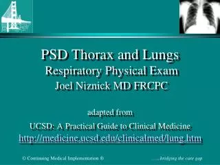
PSD Thorax and Lungs Respiratory Physical ExamJoel Niznick MD FRCPCadapted fromUCSD: A Practical Guide to Clinical Medicinehttp://medicine.ucsd.edu/clinicalmed/lung.htm
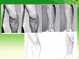
Chest configuration Pigeon chest (pectus carinatum) Barrel chest Funnel chest (pectus excavatum) Harrison’s sulcus Kyphosis Scoliosis Inspection • Cyanosis • Clubbing • Respiratory rate • Respiratory pattern • Normal • Restricted • Obstructed • Cheynes-Stokes • Painful
Differential Diagnosis of Clubbing • Cyanotic congenital heart disease • Lung disease • Cystic fibrosis • Interstitial fibrosis • Malignancy • Sarcoidosis • Bronchiectasis • Hyperthyroidism
Inspection • Rate rhythm depth effort • 14-20/min • Supraclavicular retraction and SC mastoid retraction • Posterior shape, symmetry, deformities
Palpate tactile fremitus “99,99,99” Increased, decreased, absent Resonant, dull Palpation • Lymph Nodes • Tracheal location, shift • Cutaneous lesions • Expansion • Upper lobes • Middle lobes • Posterior lobes
Contralateral Pleural effusion Hemothorax Tension pneumothorax Tracheal Deviation • Ipsilateral • Atelectasis • Fibrosis • Lung collapse • Pneumothorax
Percussion • Apices to bases • Intensity, pitch duration- resonant or dull • Diaphragmatic dullness & respiratory excursion ~ 5-6 cm • Dull: liver, spleen, heart, consolidation/collapse • Stony dull: Pleural effusion/thickening • Resonant: air filled lung • Hyper-resonant: emphysema, pneumothorax • Tympanitic: Gas filled viscus
Ohio State UniversityInteractive Guide to Physical Exam Click on image and scroll down page http://familymedicine.osu.edu/products/physicalexam/exam/
Duration Pitch Intensity Auscultation • Breath sounds • Bronchial • over sternum • Bronchovesicular • 1-2 interspace anteriorly • interscapular • Vesicular • Most of lung fields
Ohio State UniversityInteractive Guide to Physical Exam Click on image and scroll down page http://familymedicine.osu.edu/products/physicalexam/exam/
Adventitial sounds • Wheezes - continuous • Rhonchi • Crackles- intermittent • Fine • Course • Rales
Changes in voice sounds Signs of consolidation • Bronchophony “99,99,99” • Egophony “e,e,e” sounds like “ay,ay,ay” • Whispering pectorliloquay Additional sounds • Pleural rubs
Conditions • Consolidation • Collapse • Pleural effusion • Pneumothorax
Describe the Physical Signs ofLLL Pneumonia • Inspection • Palpation • Trachea • Expansion • Fremitus • Percussion • Auscultation • Broncophony • Egophony
Describe the Physical Signs ofLLL Collapse • Inspection • Palpation • Trachea • Expansion • Fremitus • Percussion • Auscultation
Describe the Physical Signs ofRight Pleural Effusion • Inspection • Palpation • Trachea • Expansion • Fremitus • Percussion • Auscultation • Whispering pectorliloquay
Describe the Physical Signs ofRight Pneumothorax • Inspection • Palpation • Trachea • Expansion • Fremitus • Percussion • Auscultation
Describe the Physical Signs ofRight Tension Pneumothorax • Inspection • Palpation • Trachea • Expansion • Fremitus • Percussion • Auscultation
COPD Clinical Features • Cough, sputum, dyspnea • Pursed lip respiration (Forced expiratory time > 6 seconds) • Hyperinflation- increased AP diameter/ hyper-resonance • Barrel chest • Reduced breath sounds • Wheezes and rhonchi • Hoover sign (paradoxical indrawing of the lateral rib margin seen during inspiration)
PulmonaryFibrosis Clinical Features • Dyspnea on exertion • Non-productive cough • Clubbing (50% in idiopathic fibrosis) • Fine bibasilar inspiratory crackles (Velcro)