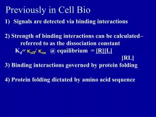
Previously in Cell Bio
This article explores the binding interactions and signaling pathways involving the Thyroid-Stimulating Hormone (TSH) and its receptor. It explains how the strength of these interactions can be quantified using the dissociation constant (Kd) and how protein folding is dictated by amino acid sequences. The piece discusses Graves' hypothesis regarding overactive thyroid responses, emphasizing the roles of T3 and T4 levels, as well as mutations in signaling processes. Additionally, it delves into membrane protein characteristics and the mechanisms by which signals are transduced across cellular membranes.

Previously in Cell Bio
E N D
Presentation Transcript
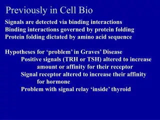
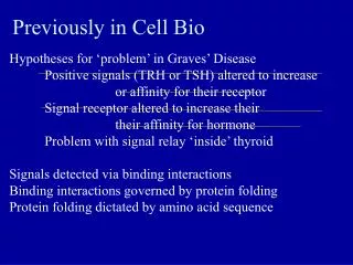
Previously in Cell Bio • Signals are detected via binding interactions • 2) Strength of binding interactions can be calculated– referred to as the dissociation constant • Kd= koff/ kon @ equilibrium = [R][L] • [RL] • 3) Binding interactions governed by protein folding • 4) Protein folding dictated by amino acid sequence
Can we predict protein structure? Motifs and Domains How do you change a protein’s shape? Alter the chain-- modifications Change the local environment– what it is floating in or binding with
TSH Receptor: What level of structure? Extracellular Plasma membrane Cytosolic TSH Receptor:from “The Thyroid Manager” Ch16
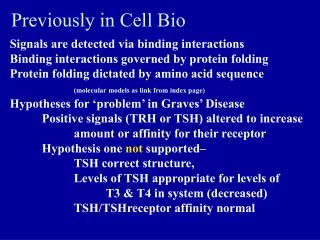
Graves’ hypothesis 1: TSH, TSH-Receptorinteraction ‘too strong’ According to this hypothesis and what we now know about protein binding…… T3 and T4 levels should be _?_ in Graves’ vs. normal. TSH levels should be __?__ in Graves’ vs. normal TSH/TSH receptor interactions should show __?___ Kd vs. normal.
Blood tests show T3 and T4 levels are elevated TSH levels are decreased TSH/TSH receptor interactions have same binding constant vs. normal. Therefore: Perfectly logical hypothesis…….
Hypothesis 2: Mutation in signaling within cell leading to increase in thyroid hormone production Normal activation is the result of signal transduction second messenger cascade How does signal transduction work? What could have gone wrong? Notsupported by data Now what?
What do we know so far? • Thyroid is ‘over reacting’ • Pituitary normally responsible for thyroid • stimulation through levels of TSH • Graves’ patients have normal/decreased levels • of TSH in blood • Binding affinity between TSH and TSH-R normal
TSH is water soluble hormone (why is this important?) More of what we know Figure 4-1. Schematic drawing of human TSH, based on a molecular homology model built on the template of a hCG model14. The a-subunit is shown as checkered, and the b-subunit as a solid line. The two hairpin loops in each subunit are marked L1, L3; each subunit has also a long loop (L2), which extends from the opposite site of the central cystine knot. The functionally important a-subunit domains are boxed. Important domains of the b-subunit are marked directly within the line drawing (crossed line, beaded line and dashed line): For further details the reader is referred to Grossman et al.2. (Reproduced from Grossman,M, Weintraub BD, SzkudlinskiMW-Endocrin Rev (4) 18:476-501,1997, with permission of the Endocrine Society). From “The Thyroid manager”
Thyroid plasma membrane is barrier • to polar molecules Even more • TSH interacts with a receptor on • the surface of thyroid cells HOW and WHY is the thyroid responding as though over-stimulated?And to get to the answer of that question: How do signals get passed across membranes?
Characteristics of Transmembrane Proteins • Hydrophobic face of protein in transmembrane region • -one continuous structure or multiple regions of 2° structure • Charges ‘anchor’ transmembrane region • Asymmetric orientation
Peripheral Membrane proteins Characteristics • Associations with membrane not as strong • Various means of attachment • -Protein-protein • -Protein-phospholipid head • - Lipid modification imbedded into membrane Fig 3-32 Molecular Cell Biology by Lodish et al. 5th ed
Membranes and membrane proteins How can a polar signal gain access to the cytosol • Direct access: From the ‘outside’ • Pores • Channels • Pumps • From cytosol to cytosol • Gap junctions
Membrane proteins Indirect access: Receptors If signaling molecule never gains access to cytosol how can the information be transmitted? Extracellular domain Plasma Membrane Cytoplasmic Domain TSH Receptor:from “The Thyroid Manager” Ch16
Transmembrane receptors • Same general structure as other transmembrane proteins • Able to bind specific ligand • Ligand binding causes conformational change What change in the TSH receptor could cause overproduction of T3 and T4 How could you test your hypothesis?
Allosteric transitions What are they, why are they important, How do they relate to signal transduction • R T state transitions • Cooperative binding Examples DNA helicase and ras (links from index page)
2nd Messengers and Signaling Cascades Getting the signal to where it needs to go For Tuesday: summarize a cascade involving. 1) cGMP 2) RTK 3) IP3 (inositol triphosphate) 4) Ca++ 5) DAG (diacyl glycerol) Email me one paragraph summary of how that one cascade works by midnight Monday (think ‘handout’)