Download

1 / 36
380 likes | 1.14k Vues
Cell Reproduction or How cells make copies of themselves. Also called Cell Division. Cell Division. Cell division consists of two phases: 1. the division of the nucleus, and 2. the division of the cytoplasm (cytokinesis). 2 Kinds of Nuclear Division.

E N D
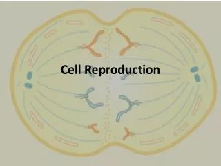
Cell ReproductionorHow cells make copies of themselves Also called Cell Division
Cell Division • Cell division consists of two phases: • 1. the division of the nucleus, and • 2. the division of the cytoplasm (cytokinesis)
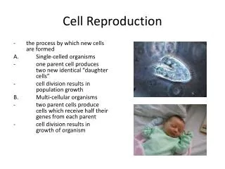
2 Kinds of Nuclear Division • 1. Mitosis – mitosis divides the nucleus so that both resulting new cells (daughter cells) are genetically identical…Same amount of DNA • 2. Meiosis – meiosis produces daughter cells that contain half the genetic information… half the amount of DNA.
Before Division Begins • Before a cell can successfully divide, the DNA must be packaged so it does not get damaged. The “stringy” form of DNA (chromatin) is coiled into structures called “chromosomes”.
Each Chromosome is made up of two identical halves called “sister chromatids” joined at the centromere. Each Chromatid is a single, coiled DNA molecule. The point where two sister chromatids are connected. Sister Chromatids
Diploid Cells(symbolized as 2n) • Diploid cells (2n) have 2 copies of every chromosome, forming what is called a “Homologous” chromosome pair. In Diploid cells, one pair of chromosomes comes from the mother and the other pair comes from the father. The diploid number for humans is 46, or 2n = 46,…. Or you could say…..Humans have 23 homologous pairs, or…. Humans have 92 chromatids.
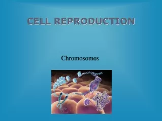
Chromosomes Chromosomes
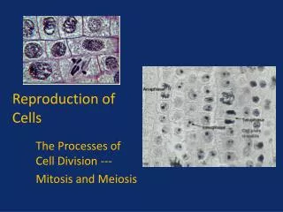
The Life Cycle of a cell is called The “Cell Cycle” The Cell Cycle consists of 5 Phases 1.Interphase (part of the cell cycle, but not part of mitosis) 2.Prophase 3.Metaphase 4.Anaphase 5.Telophase These 4 phases are known collectively as “Mitosis”
Interphase 90% of cell’s life is spent in interphase During Interphase the cell grows, duplicates its chromosomes and performs its normal job. Interphase has 3 stages The Events of Interphase G1 stage - first gap - Cell grows and carries out regular biochemical functions. S stage – synthesis - DNA is replicated or synthesized. G2 stage - second gap - Cell completes preparations for division…..a cell can complete S, but fail to enter G2.
nuclear envelope Plant Cells Animal Cells
Mitosis(the splitting of the nucleus)prophasemetaphaseanaphase telophase
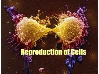
Mitosis • A cell that has grown in size and is about to divide is called a “Mother Cell”. • As a result of Mitosis and cytokinesis the Mother cell splits into two genetically identical “Daughter Cells”. Mother Cell
The Events of Prophase • Nucleoli disappear. • Chromatin condenses into the chromosomes. • Nuclear Envelope dissolves… the nucleus comes apart • Centrioles (MTOC’s) separate and move to opposite ends of the cell. • Microtubules from each MTOC connect to a specialized region of the centromere called the kinetochore. This moves the chromosomes back and forth. • Mitotic spindle begins to form.
Plant Cells Animal Cells
Events of Metaphase • Chromosomes line up at the equator of the cell…. called the “metaphase plane”. • Centrioles arrive at opposite ends of the cell. • Spindle apparatus fully developed. • Metaphase ends when the microtubules pull each chromosome apart into two chromatids. Once separated from its sister chromatid, each chromatid is now called a chromosome. To count the number of chromosomes, at any time, count the number of centromeres. centriole centriole metaphase plane
Plant Cells Animal Cells
Events of Anaphase Chromosomes • Anaphase begins when the chromosomes separate. • Microtubules shorten as tubulin units uncouple, the chromosomes are pulled away from each other toward opposite ends of the cell. • Cell elongates; poles move slightly further apart. • Anaphase ends when the chromosomes reach their respective ends of the cell.
Plant Cells Animal Cells
Events of Telophase • Chromosomes uncoil back to chromatin. • Nuclear envelope reforms. The nucleus reforms in each newly formed cell. • Nucleoli reappear. • Spindle fibers disappear. • Simultaneously Cytokinesis usually starts.
Plant Cells Animal Cells
Animal Cell Cytokinesis • “Cleavage furrow” forms. • Microfilaments contracts and divides the cytoplasm into two parts.
Plant Cell Cytokinesis • Cell plate develops from Golgi vesicles. • New cell wall developed around the cell plate. p
Regulation of Cell Division • Must be controlled. • Rate of cell division depends on the cell type. • Example: skin cells divide frequently liver cells divide as needed brain cells rarely or never divide Cells will stop dividing when the surrounding cell density reaches a specific level….... this is called “Density-Dependent Inhibition” • When density is high - no cell division. • When density is low - cells divide.
Cancer Cells • A tumor is a large mass of cells. Tumors form because cells do not stop dividing. The control mechanisms for cell division have failed.
Why Cells Divide • As cells grow in size they become less healthy.... If they don’t divide (split in two) they will die.
2 Reasons Why Cells Become Less Healthy • 1. Surface area-to-Volume ratio becomes too small. • 2. The nucleus is limited in regulating cell activities…. its genome-to-volume ratio becomes too small.
Surface area-to-Volume Ratio • When a cell grows, the volume of the cell increases faster than the surface area surrounding it. • When we say “the surface area-to-volume ratio is large”, that means there is a large surface area relative to volume. • When we say “the surface area-to-volume ratio is small”, that means the surface area is small relative to volume. • When the surface-to-volume ratio is large, the cell can effectively react with the outside environment…..for example…. adequate amounts of water and oxygen can move into the cell, and wastes can rapidly be eliminated. • When the surface-to-volume ratio is small the cell is unable to exchange enough substances to service the cell. The cell dies.
Genome-to-Volume Ratio • The DNA in a cell is referred to as its “genome” (all of its genes). • The genome controls all of the cell’s activities by producing enzymes…. which trigger and control the cell’s necessary functions. • Because there is a finite amount of DNA, the amount of enzymes is limited. • As the cell grows the volume increases, but the amount of DNA remains the same…. In other words “the genome-to-volume ratio” decreases….. Eventually there is not enough DNA to regulate the cell….. cell functions decrease and the cell dies.