Download
1 / 17
170 likes | 198 Vues
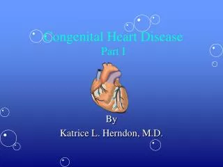
TREATMENT OF CONGENITAL HEART DISEASE BASIS OF TREATMENT OF ASD, VSD &PDA. DR AFTAB YUNUS FRCSEd. CHAIRMAN CARDIAC SURGERY KING EDWARD MEDICAL UNIVERSITY, LAHORE. CONGENITAL HEART DISEASES. Acyanotic Atrial Septal Defects. Ventricular Septal Defects. Patent Ductus Arteriosus. Cyanotic

E N D
TREATMENT OF CONGENITAL HEART DISEASEBASIS OF TREATMENT OF ASD, VSD &PDA DR AFTAB YUNUS FRCSEd. CHAIRMAN CARDIAC SURGERY KING EDWARD MEDICAL UNIVERSITY, LAHORE.
CONGENITAL HEART DISEASES • Acyanotic • Atrial Septal Defects. • Ventricular Septal Defects. • Patent Ductus Arteriosus. • Cyanotic • Fallot’s Tetralogy. • Total Anomalous Venous Drainage. • Transposition of Great Arteries.
CONGENITAL HEART DISEASES • Atrial Septal Defects • Ventricular Septal Defects. • Patent Ductus Arteriosus.
ATRIAL SEPTAL DEFECTS. • Defect (Hole) in inter atrial septum. • Blood flows from left atrium to right atrium. • Blood flow through pulmonary (Lungs) circulation increases. • Changes in Pulmonary (Lungs) blood vessels.
Ventricular septal defects • Defect (Hole) in the inter-ventricular septum. • Blood flow from left ventricle to right ventricle. • Blood flow through pulmonary (Lungs) circulation increases. • Changes in pulmonary (Lungs) blood vessels
Patent ductus arteriosus • Persistence of fetal structure Ductus arteriosus. • Connects Aorta with left pulmonary artery. • Blood flows from aorta to left pulmonary artery. • Lungs blood flow increases. • Changes in pulmonary (Lungs) blood vessels
treatment • To reduce the blood flow to lungs to normal. • Closure of these abnormal blood flow channels. • With time. • By cardiac physician • By cardiac surgeon
Closure with time • Observe for 1 year. • if no symptoms. • In Premature infants. • Drugs. (PDA: NSAID ibuprofen/indomethacin)
Closure by cardiac physician • Not open heart surgery. • Through femoral artery or vein. • Place a device to close the defect.
Closure by cardiac surgeon pda • Left postero-lateral thoracotomy. • PDA dissected and doubly ligated. • Thoracotomy closed.
Closure by cardiac surgeon asd • Median Sternotomy. • Heart-Lung Machine. • Right atrium opened. • Hole in septum either closed directly or by a patch. • Off Heart-Lung Machine. • Closure
Closure by cardiac surgeon Vsd • Median Sternotomy. • Heart-Lung Machine. • Right atrium opened. • Hole in septum closed by a patch. • Off Heart-Lung Machine. • Closure