Download
1 / 51
510 likes | 748 Vues
Automatic Volumetric Breast Density Assessment in the eXtensible Imaging Platform. Rachel Embree and Christina Sillery David Channin, MD, Advisor In Cooperation with Alex Shnayder, Pat Mongkolwat, Ray Wu. Overview. Introduction Material and Methods Results Discussion Conclusion.

E N D
Automatic Volumetric Breast Density Assessment in the eXtensible Imaging Platform Rachel Embree and Christina Sillery David Channin, MD, Advisor In Cooperation with Alex Shnayder, Pat Mongkolwat, Ray Wu
Overview • Introduction • Material and Methods • Results • Discussion • Conclusion
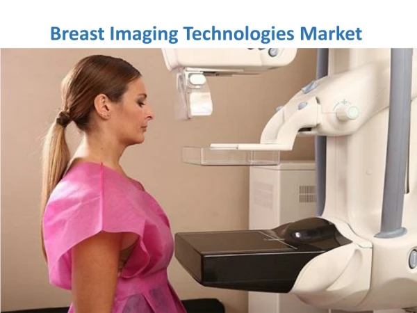
What is Breast Density? • The breast is composed of fibroglandular tissue embedded in a background of fatty tissue. The amount of fibroglandular tissue and fat varies among women. • Breast cancer arises in this fibroglandular tissue. • Mammography is projection radiography of the breast. • In mammograms, dense fibroglandular tissue attenuates more radiation than does fat. • Breast density refers to the appearance of this fibroglandular tissue in a mammogram [5]
Breast Density • Positively correlated with breast cancer • Four to six times greater risk of breast cancer for women with greater than 60% dense tissue [1] • Breast density as a risk factor accounts for as many as 30% of breast cancer cases [1] • Can be changed by hormonal, dietary and other interventions
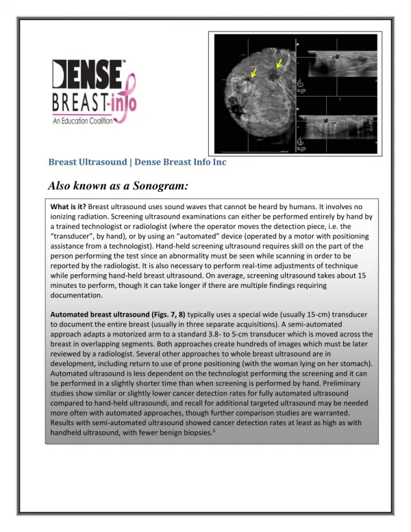
Assessment of Breast Density in Mammograms • Subjective Visual Assessment by Radiologists (Mandatory) • Score the breast on a 4-point scale in the Breast Imaging-Reporting and Data System (BIRADS), based on patterns developed by Wolfe [5] • Manual Image Processing Techniques (for research) • Planimetry • Interactive Thresholding
Previous Approaches • Interactive Thresholding
Goal • Create a breast density assessment program that is… • Automated • Volumetric • Accurate • In the eXtensible Imaging Platform (XIP)
What is XIP? • eXtensible Imaging Platform • Funded by NIH, NCI Cancer Bioinformatics Grid (caBIG) Program • A program to share data, informatics tools, technologies and infrastructure between the funded Comprehensive Cancer Centers across the nation • Open Source, Open Standards, Open Architecture • Platform for development of medical image processing and analysis applications for research and clinical purposes. • Will support the DICOM Application Hosting standard to facilitate transportability of XIP applications
XIPBuilder • Visual Programming Environment for developing imaging applications; based on OpenInventor. • Contains modules (lots!) that can be assembled to perform sophisticated tasks. • Includes • vTK – The Visualization Toolkit (NIH) • iTK – The Insight Toolkit for Registration and Segmentation (NIH) • Extensible by creating new plug-n-play modules
Algorithmic Foundations • Highnam et al 1997 • Standard Mammogram Form (SMF) • Volumetric approach • Mammograms on X-ray film • Van Engeland et al 2006 • Improved upon SMF • Full Field Digital Mammography (FFDM) • Utilized DICOM headers
The Model [2]
Volumetric • Based on Van Engeland’s algorithm [4] • Tissue composition is computed at each pixel and represents a rectangular cylinder of tissue • Area of the pixel is computed from detector characteristics and the geometry of acquisition. • Height of the rectangular cylinder is computed from the compression thickness.
SoXIPLoadDICOM • Loads … • DICOM Image Pixel Data • Necessary DICOM Header Information
Input Output SoItkBinaryThresholdImageFilter • Each pixel compared to threshold value • Output image of 0 and 1 values
Input 2 Input 1 Output SoItkMultiplyImageFilter • Pixel-wise multiplication of two images • Lays background mask on top of original image
SoXIPLinearAttenuationCoefficients • Determines difference between effective attenuation of dense tissue and fat • This Attenuation Difference Coefficient is chosen from a table [4] • Based on DICOM header information • Anode target material • Filter material • kVp • Breast Thickness
SoXIPBreastDenseTissueVolumeCalculation • hd(r) : height of dense tissue at pixel r • µd, eff - µf, eff : Attenuation Difference Coefficient • g(r) : current pixel value • gf : fatty tissue reference value [4]
SoXIPBreastDenseTissueVolumeCalculation • hd(r) : height of dense tissue at pixel r • µd, eff - µf, eff : Attenuation Difference Coefficient • g(r) : current pixel value • gf : fatty tissue reference value [4]
SoXIPBreastDenseTissueVolumeCalculation • hd(r) : height of dense tissue at pixel r • µd, eff - µf, eff : Attenuation Difference Coefficient • g(r) : current pixel value • gf : fatty tissue reference value [4]
SoXIPBreastDenseTissueVolumeCalculation • hd(r) : height of dense tissue at pixel r • µd, eff - µf, eff : Attenuation Difference Coefficient • g(r) : current pixel value • gf : fatty tissue reference value [4]
SoXIPBreastDenseTissueVolumeCalculation • hd(r) : height of dense tissue at pixel r • µd, eff - µf, eff : Attenuation Difference Coefficient • g(r) : current pixel value • gf : fatty tissue reference value [4]
Where does fatty tissue reference value, gf , come from? • Maximum pixel value (minimum attentuation) in the interior of the breast (this should represent a pure fat pixel). • Calculate a histogram of the image. • Select the pixel value at 80% (empirical determination) of the maximum pixel values (avoids skin edges; low attenuation due to incomplete thickness).
Input Output SoItkBinaryThresholdImageFilter • Adjust parameters to detect breast edge instead of entire region
SoXIPBreastTotalTissueVolumeCalculation • h(r) : total height of tissue at pixel r • H : compressed breast thickness • d(r) : Euclidean distance to the edge between breast tissue and background [4]
SoXIPBreastTotalTissueVolumeCalculation • h(r) : total height of tissue at pixel r • H : compressed breast thickness • d(r) : Euclidean distance to the edge between breast tissue and background [4]
SoXIPBreastTotalTissueVolumeCalculation • h(r) : total height of tissue at pixel r • H : compressed breast thickness • d(r) : Euclidean distance to the edge between breast tissue and background [4]
SoXIPBreastTotalTissueVolumeCalculation • h(r) : total height of tissue at pixel r • H : compressed breast thickness • d(r) : Euclidean distance to the edge between breast tissue and background [4]
SoXIPBreastTotalTissueVolumeCalculation • If Euclidean distance is less than half of breast compression … • use first formula in calculation of total tissue volume [4]
SoXIPBreastTotalTissueVolumeCalculation • If Euclidean distance is greater than half of breast compression… • use breast compression in calculation of total tissue volume [4]
x 100 = Percent Density Calculator • Module in XIPBuilder Dense Tissue Volume Total Tissue Volume
Separator • Module in RADBuilder • Displays output • In this case, text
Methods II • 20 CC full-field digital mammograms • Prior manual breast density assessment using ImageJ • Determine breast density using Cumulus™, a popular interactive thresholding program • Determine breast density with the automated XIP solution
Methods III • Compare the three measurements of breast density to determine Kendall’s Coefficient of Concordance • Use that coefficient to determine a χ2 that allows testing of the null hypothesis: There is no agreement in the assessment of breast density by the three methods
Results • The overall Kendall’s Coefficient comparing the three systems was 0.502 • For 20 cases and three systems, the χ2 is 57.276 which allows the rejection of the null hypothesis (χ2 of 30.14, 19 degrees of freedom, α =0.05).
Conclusion • It was possible to develop, in XIP, an automatic software application to measure volumetric breast density in mammograms. • The automatic measurement agreed well with two, independent, manual thresholding based techniques in common, current use.
Future Work • MLO view capability • Anisotropic Filter • Improve fatty reference value identification • Validate this and other techniques against ground truth (MRI)