Download
1 / 1
10 likes | 224 Vues
Silicon detectors for combined MR-PET and MR-SPECT imaging.

E N D
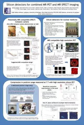
Silicon detectors for combined MR-PET and MR-SPECT imaging Andrej Studen, Karol Brzezinski, EnricoChesi, Vladimir Cindro, Neal H. Clinthorne, Milan Grkovski, Borut Grošičar, Klaus Honscheid, S. S. Huh, Harris Kagan, Carlos Lacasta, Gabriela Llosá , Marko Mikuž , Vera Stankova, Peter Weilhammerand Dejan Žontar Jožef Stefan Institute, Ljubljana, University of Michigan, Ohio State University, IFIC/CSIC-UVEG Valencia, University of Ljubljana Email: andrej.studen@ijs.si Potentially MRI compatible SPECT: Compton camera Silicon detectors for nuclear medicine • Adjusted pad (pixel) detectors with characteristics: • Resistivity > 100 kOhmcm • Thickness of 1 mm • P+nn+ structure formed with planar processing • P+ side segmented to square pads, 1.4 mm sized (1 mm under test) • Size of 4.4 by 2.2 cm2, 512 pads • Readout with parallel amplifiers, 128 per ASIC • ASIC: VATAGP series by GM-IDEAS (ver. 3 and 7) • 150 ns fast shaper for trigger, 500 ns -3 us slow shaper for energy • Insensitive to magnetic fields • Compact, flexible, sturdy. 1.4 x 1.4 mm2 (Non MRI) Compton camera demonstrator at UoM Silicon detector combined with a standard (ADAC) gamma camera without a collimator. Collimator only used to compress imaging field to a single slice. 99mTc 140 keV Reconstruction based on Compton kinematics tracked by impact position measurement in both sensors and energy measured in the silicon scatterer. Different sources used. MRI compatible high resolution PET Single slice PET 131I 364 keV Volume PET 22Na 511 keV Photograph of the inner ring make-up in single slice mode. The object is placed on a rotary table. Lead collimators used to filter the image slice. Based on the dual ring efforts. The inner ring is silicon based & MRI compatible. Tested in single slice mode with F-18 filled high-resolution Derenzo phantom (rods 1.2 mm to 4.8 mm). Number of collected counts indicated, MLEM reconstruction used for 60 angular views. Tested with Na-22 point sources in volumetric mode. No septa, 60 angular views. Suggested MRI compatible design The scatterer could be used as a probe within the MRI bore with external gamma camera placed in the low field environment. Cochran, E et al. Performance of electronically collimated SPECT imaging system in the energy range from 140 keV to 511 keV. 2008 NSS Conf Rec: 4618-4621, 2008. N.H. Clinthorne et al. Silicon as an unconventional detector in positron emission tomography, accepted to NuclInst Meth A: 10.1016/j.nima.2012.05.026. Compression in positron range measured at 7 T with high resolution silicon PET B 68Ga - e+ 1.9 MeV A dedicated positron range setup To test beneficial positron range compression in high magnetic field. Silicon detectors combined with MRI compatible mechanics were placed in 7 T large bore MRI magnet at OSU. No degradation in performance due to magnetic field was observed. Detectors were separated by 17 cm , the sources were placed on a pneumatically driven rotary table with 2 degree reference marks. Beneficial resolution improvement in the axial plane. • Lorentz force acting on the passing positron curves its path so it annihilates closer to the creation point. • Tested with higher energy positron emitters (94mTc, 68Ga) where effect is more pronounced. Out of plane artifacts & multiple field orientation Effect is highly directional, in orientation along the field, no compression is attained. Multiple orientations restore symmetry of the point spread function. Artifacts from out of plane sources alleviated by accounting for positron range in MLEM reconstruction. Off plane offset 0 T 7 T 3 mm 2 mm 1 mm 2D 3D D. Burdette et al. A device to measure the effects of strong magnetic fields on the image resolution of PET scanners, NuclInst Meth A 609 (2–3): 263-271, 2009.