The Occipital Lobe
140 likes | 486 Vues
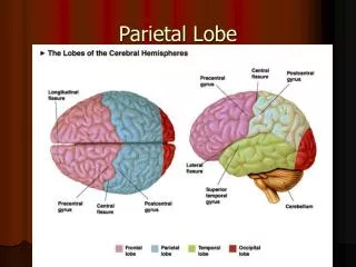
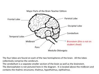
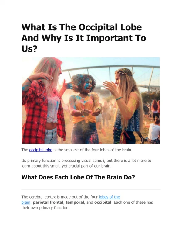
The Occipital Lobe. “The Occi”. By: Bri Vf. Lindsey W. Jarred T. The Occipital Lobe is the rearmost lobe in each cerebral hemisphere of the brain. It contains the visual center of the brain. - It is one of the main lobes/regions of the cerebral cortex

The Occipital Lobe
E N D
Presentation Transcript
The Occipital Lobe “The Occi” By: Bri Vf. Lindsey W. Jarred T.
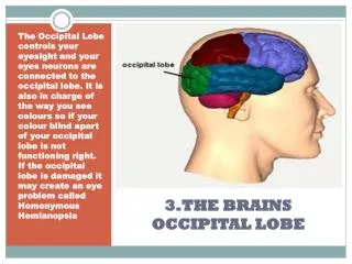
The Occipital Lobe is the rearmost lobe in each cerebral hemisphere of the brain. It contains the visual center of the brain. • - It is one of the main lobes/regions of the cerebral cortex • Main visual processing (color and face recognition) • Occipital Cortex-(anatomy) a somewhat rounded subdivision of a bodily organ or part. Notes and Vocabulary
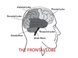
What body part does it control? The occipital lobe is located at the back of our brain. It is responsible for receiving and processing visual information from our eyes.
What happens when this is injured? They are not particularly vulnerable to injury because of their location at the back of the brain, although any significant trauma to the brain could produce subtle changes to our visual-perceptual system, such as visual field defects and scotomas.
Visual Hallucination(Epilepsy) Seizures can occur in the occipital lobe, which can cause visual hallucinations. The patient may see rapid blinking, colored lights or flickering lights. Occipital lobe seizures, which the NYU Comprehensive Epilepsy Center states account for 5 to 10 percent of epilepsy cases, can be triggered by flashing lights.
Treatment for Epilepsy The majority of epileptic seizures are controlled by medication, particularly anticonvulsant drugs(pain killers). The type of treatment depends on the person’s frequency of seizures, a persons age and weight, overall health, and medical history. Drugs Used to treat Epilepsy- Dilantin or Phenytek Phenobarbital Tegretol or Carbatrol Mysoline Zarontin Depakene Depakote, Depakote ER Valium and similar tranquilizers, such as Tranxene and Klonopin Newer drugs to treat epilepsy include: Felbatol Gabitril Keppra Lamictal Lyrica Neurontin Topamax Trileptal Zonegran
Does Lincoln’s face look normal? Activity! Does Lincoln’s face look normal?
It seems normal but now, look at it upright: Lincoln’s eyes do not look quite right! It seems normal but now, look at it upright: Lincoln’s eyes do not look quite right! Some neurons in the brain seem specialized in processing faces. Faces are usually seen upright. When presented upside down, the brain no longer recognizes a picture of a face as a face but rather as an object. Neurons processing objects are different from those processing faces and not as specialized. As a consequence these neurons do not respond to face distortions as well. This explains why we miss the weird eyes when the face is inverted.
Another great example of an illusory contour! The baby’s head is on the left, the baby’s feet are against the trunk of the tree on the right.
Can you put the fish in the fishbowl? Stare at the yellow stripe in the middle of the fish in the picture below for about 10–20 sec. Then move your gaze to the fish bowl.
Did you see a fish of a different color in the bowl? You have just experienced an afterimage.In the retina of your eyes, there are three types of color receptors (cones) that are most sensitive to either red, blue or green. When you stare at a particular color for too long, these receptors get “fatigued.” When you then look at a different background, the receptors that are tired do not work as well. Therefore, the information from all of the different color receptors is not in balance. This will create the color “afterimages.”
Quiz 1.What does the occipital lobe control and where is it located? 2.What part of the body does it operate?