Download
1 / 1
10 likes | 119 Vues
Quantitative Volume and Density Response Assessment: Sarcoma and HCC as a Model. Lawrence H Schwartz, M.D. and Binsheng Zhao, D.Sc. Columbia University Medical Center, New York. Objective.

E N D
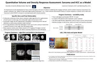
Quantitative Volume and Density Response Assessment: Sarcoma and HCC as a Model Lawrence H Schwartz, M.D. and Binsheng Zhao, D.Sc. Columbia University Medical Center, New York Objective A critical barrier to the development of new cancer therapies is the lack of evidence validating the imaging methods used for patient response assessment. Our project evaluates the ability of improved image analysis techniques, e.g., tumor volume and necrosis fraction, to assess response in cancers such as HCC and Sarcoma in which RECIST is known to correlate poorly with clinical outcome. Progress Summary – variability study Specific Aims and Final Deliverables • Three radiologists participated; RECIST 1.0 used • A subset of 30 patients with metastatic cancer selected • The three segmentation algorithms used to assist in tumor measurement • Relative changes in total tumor volume at 6-wk from baseline calculated • To develop and deliver three robust computer-aided algorithms for segmentation • of solid tumors in the lungs, liver and lymph nodes and necrosis in HCC • To provide insight into the magnitude of variability in measuring tumor volume • (diameter as well) using computer-aided methods • A CT image dataset containing radiologists’ mark-up of tumor contours made from • a subset of tumors in the lungs, liver and lymph nodes • New response metrics and criteria, based on CT imaging of changes in tumor • volume and necrosis fraction, for better assessing response to novel therapies Upper 95% limit Lower 95% limit R1 / R2 R1 / R3 Inter readers R2 / R3 Progress Summary – algorithm and platform development U01 / R01 Hub and Spoke Model Segmentation algorithms developed for lesions in the lungs, liver and lymph nodes Significant differences are marked in red Effects of CT slice thickness and reconstruction algorithm on volume measurement – a phantom study Publications Tan Y, Schwartz LH and Zhao B, Segmentation of lung tumors on CT scans using watershed and active contours. Med Phys. 2013, Apr; 40(4):043502. doi: 10.1118/1.4793409. Zhao B, Lee S, Lee HJ, Tan Y, Qi J, Persigehl T, Tan T and Schwartz H,Variability in assessing treatment response: metastatic colorectal cancer as a paradigm. CCR (accepted). 3. Multiple abstracts resulted from this study have been published. Open source imaging platform with integrated tumor segmentation algorithms