Download
1 / 176
1.77k likes | 1.93k Vues
15. The Lymphatic and Immune Systems: Your Defense Systems. Multimedia Asset Directory. Slide 18 Lymphatic Drainage Massage Video Slide 23 Lymphatic System Animation Slide 40 Proper Hand Washing and Gloving Techniques Video Slide 69 Fever Animation Slide 112 Leukemia Video

E N D
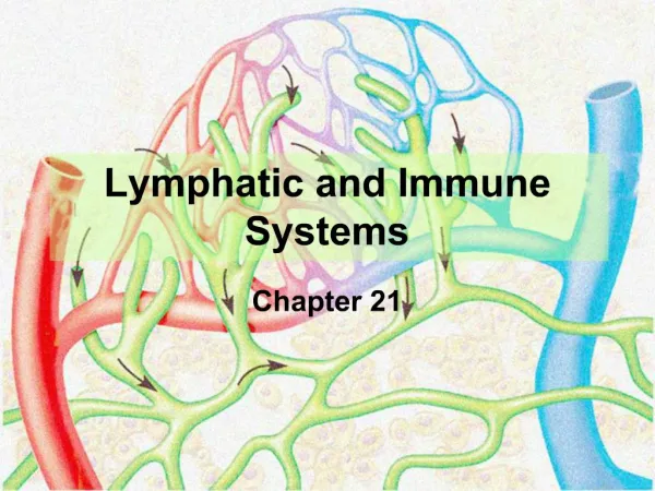
15 The Lymphatic and Immune Systems: Your Defense Systems
Multimedia Asset Directory Slide 18 Lymphatic Drainage Massage Video Slide 23 Lymphatic System Animation Slide 40 Proper Hand Washing and Gloving Techniques Video Slide 69 Fever Animation Slide 112 Leukemia Video Slide 132 Allergic Rhinitis Video Slide 133 EpiPen Video Slide 134 Anaphylaxis Animation Slide 135 Pharmacy Video Slide 136 Nuclear Medicine Video
Introduction • The immune and lymphatic systems work to protect your body from pathogens that can produce disease. • Without these systems, you would not survive for long.
Learning Objectives • List and describe the major components of the immune system and their function(s). • Explain the antigen–antibody relationship. • Name and describe the functions of the blood cells responsible for protecting the body from invasion. • Discuss how inflammatory responses and fevers relate to infection.
Learning Objectives • Compare innate immunity to adaptive immunity. • Describe the function of lymphocytes and helper cells in the immune response. • List and describe several common diseases of the immune system.
basophils (BAY soh filz) cytokines (SIGH tow kines) cytotoxic T cells (SIGH tow TOX ick) dendritic cells (Den DRIH tick) eosinophils (EE oh SIN oh filz) histamine (HISS tuh meen) interferon (in ter FEAR on) interleukins (IN ter LOO kinz) leukocytes (LOO koh sights) Pronunciation Guide Click on the megaphone icon before each item to hear the pronunciation.
lymph nodes (LIMF nohdz) lymphocytes (LIMF oh sights) macrophages (MAC roh fage ez) neutrophils (NOO troh fill) thoracic duct (thoh RASS ik) thymus (THIGH mus) tumor necrosis factor (neh KROH sis) Pronunciation Guide Click on the megaphone icon before each item to hear the pronunciation.
The Defense Zone • If a group of pathogens try to enter your body, they must first get past the barriers, such as intact skin and the secretions of your mucous membranes. • If the pathogen does get into the body it is recognized as NOT belonging. • This stimulates a series of responses to neutralize the pathogen.
The Defense Zone • Weapons include the special cells and powerful chemicals of the immune and lymph systems. • Chemicals stimulate inflammatory and clean-up responses.
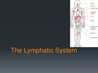
The Lymphatic System • The lymphatic system is both the transport system and barracks of the immune system. • It works closely with the cardiovascular system. The lymphatic system has four functions: • Recycling fluids lost from the cardiovascular system • Transporting pathogens to the lymph nodes where they can be destroyed
The Lymphatic System • It works closely with the cardiovascular system. The lymphatic system has four functions: • Storage and maturation of some types of white cells • Absorption of glycerol and fatty acids from food you eat
Structures of theLymphatic System • The smallest pipes of the lymphatic system are called lymph capillaries, and they run parallel to your blood capillaries. • Lymph capillaries form a network between cells of connective tissues. • The fluid filling the lymph capillaries is called lymphatic fluid, or lymph.
Structures of theLymphatic System • Lymph capillaries empty into lymphatic vessels, which are similar to veins including valves. Body movement and contraction of smooth muscles propels lymph through the system.
Figure 15-1 Relationship between blood and lymph capillaries.
Lymph Nodes • Large vessels empty into lymph nodes, ranging in size from an olive to a pin head. Many lymph vessels empty into each node, ensuring all lymph fluid passes through. • Lymph nodes are small, encapsulated bodies divided into sections. • Nodes consist of sections of lymphatic tissue containing WBCs known as lymphocytes.
Lymph Nodes • The lymphatic tissue is surrounded by lymphatic sinuses filled with lymph fluid. • Lymph nodes filter and destroy pathogens using the WBCs. • Lymph nodes are concentrated in the cervical, axillary, inguinal, pelvic, abdominal, thoracic, and supratrochlear areas. Adenoids and tonsils, the spleen, and the thymus also contain lymphatic tissue.
Click here to view a video on the topic of Lymphatic Drainage Massage. Back to Directory
Lymphatic Trunks • Lymphatic vessels exiting lymph nodes empty into one of several lymphatic trunks. • These trunks, named for their location, include the lumbar, intestinal, intercostal, bronchomediastinal, subclavian, and jugular trunks. • Lymphatic trunks empty into one of two collecting ducts.
Collecting Ducts • The lumbar, intestinal, and intercostal trunk all empty into the thoracic duct – a large duct that runs from the abdomen up through the diaphragm and into the left subclavian vein. More than 2/3 of your lymphatic system drains into this duct. • The bronchomediastinal, subclavian, and jugular trunks empty into the right lymphatic duct, a smaller duct within the right thorax which empties into the right subclavian vein.
Circulation ofLymphatic Fluid • The circulation of lymphatic fluid follows this pattern: • Blood to tissue • Tissue to lymphatic capillaries • Lymphatic capillaries to lymphatic vessels • Lymphatic vessels to lymph nodes • Lymph nodes to lymphatic vessels • Lymphatic vessels to lymphatic trunks • Lymphatic trunks to collecting ducts • Collecting ducts to subclavian veins, re-entering the blood stream
Click here to view an animation on the topic of the Lymphatic System.The animation may take a moment before playing. Back to Directory
The Spleen • The spleen is a spongy organ in the upper left quadrant of the abdomen, structurally similar to lymph nodes but instead of lymphatic sinuses the spleen has blood sinuses. • Surrounding the blood sinuses are islands of white pulp containing lymphocytes and islands of red pulp containing both RBCs and WBCs.
The Spleen • The spleen functions to remove and destroy old, damaged, or fragile RBCs. • The spleen also filters pathogens from the blood stream and destroys them like the lymph node. • It is not an essential organ and can be surgically removed. Removal in children can severely compromise immunity, but has far less of an effect on adults.
The Thymus • The thymus is a soft organ located between the aortic arch and sternum. • The thymus is very large in children because of all the new infections it must fend off. • It gets smaller or even disappears in adults as the immune system fully matures in its ability to fight infection.
The Thymus • The thymus is packed with lymphocytes, which mature into a type of WBC called a T-lymphocyte. • The thymus also secretes a hormone that stimulates maturation of the T-lymphocyte in lymph nodes.
Clinical Application:Cancer Stages • Cancer is staged and prognosis is determined by the amount of metastasis. The lymphatic system is amazingly efficient at capturing and transporting pathogens from connective tissue to lymph nodes for destruction. • Unfortunately, this can also be a liability when cancerous cells develop because they can hitch a ride with lymph fluid to distant parts of the body.
Clinical Application:Cancer Stages • Metastasized cancers are more likely to be fatal. Early detection of cancer improves outcome. • Stage 1 cancer has no spread from origin. • Stage 2 has spread to nearby tissue. • Stage 3 has spread to nearby lymph nodes. • Stage 4 cancer has spread to distant locations and is often terminal.
The Immune System • The immune system provides the weapons, and the troops, to protect your body from invasion. • The immune system is comprised of cells, chemicals, and barriers that protect your body from pathogens. • Some processes are active, some passive, some inborn, and others change with experience.
Antigens • Cells have molecules on the outer surface of the membrane to identify them as friend or foe. These molecules are antigens. • Each living thing has unique cell surface antigens, allowing your immune system to distinguish between your own cells and invaders. This ability is called self vs. non-self recognition and is the heart of how the immune system functions. • A well-functioning immune system ignores your self antigens and attacks non-self antigens.
Antibodies • Your body makes proteins that bind to antigens, eventually destroying them. • These proteins are called antibodies and are one of the most potent weapons of the immune system. • Antibodies are called into action when a foreign antigen invades the body.
Innate Immunity • Innate immunity is the front line of defense against invasion. • Innate immunity is your body’s inborn ability to fight infection. • Innate immunity prevents invasion, or if the invasion occurs, it takes steps to prevent the spread of infection.
Innate Immunity • Innate immunity can only recognize that something is not you, it can’t actually identify the invader. • Innate immunity is a collection of crude mechanisms, like weapons of mass destruction, killing pathogens and healthy tissue alike.
Adaptive Immunity • Your innate immunity is backed up by adaptive immunity that specifically targets invaders and spares healthy tissue as much as possible. • Adaptive immunity remembers invaders from previous encounters and prepares for future invasion, improving the response with experience by learning and changing.
Adaptive Immunity • Innate and adaptive immunity are not two separate entities, but work together, with innate immunity preparing the way for adaptive immunity, weakening some pathogens and stimulating components of adaptive immunity. Adaptive immunity, in turn, stimulates innate immunity attacking the pathogen on two fronts.
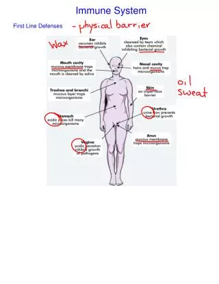
Components of the Immune System • Physical barriers exist to act as a first line of defense. • White blood cells are a second line of defense against pathogens that manage to get by the barriers. • There are numerous chemicals that play a role in destroying pathogens. • Finally, body processes kick in to help fight infection.
Physical Barriers • Anything that prevents invaders from getting inside your body prevents infection. • Physical barriers are located in areas most likely to be invaded. • Skin, and the mucous membranes of the eyes, digestive, respiratory, and reproductive systems act as barriers.
Physical Barriers • Not only are these surfaces difficult to penetrate, they are packed with WBCs and lymph capillaries to trap invaders. • The fluids associated with these physical barriers, including tears, saliva, urine, mucous secretions, and sweat, all contain chemicals that act as barriers. • Barriers are part of innate immunity.
Click here to view a video on the topic of Proper Hand Washing and Gloving Techniques. Back to Directory
White Blood Cells • WBCs (leukocytes) are the cells responsible for defending your body against invaders. • WBCs, after being born in the bone marrow, move to other parts of the body to grow and mature until they are needed during an invasion. • WBCs are not released into the blood stream until they are needed.
Classification of Leukocytes • There are many types of leukocytes. • Leukocytes are divided into two groups: • Polymorphonuclear granulocytes are cells with granules, or spots, in their cytoplasm. • Mononuclear agranulocytes have no granules in their cytoplasm.
Neutrophils • The body has various types of WBCs that are called upon in different circumstances. • Neutrophils are granulocytes whose function is phagocytosis, ingesting pathogens and cellular debris. • Neutrophils are born in the bone marrow and are the most common leukocyte in the blood stream.
Neutrophils • Neutrophils are the first responders at the site of invasion, cleaning up the area by ingesting pathogens and releasing chemicals which increase tissue damage and inflammation, stimulating immune response. • Neutrophils are part of innate immunity.
Macrophages • Macrophages are modified monocytes which leave the blood stream and enter tissues. • They are also phagocytic, active in the later stages of an infection. • These cells release chemicals which stimulate the immune system. • Macrophages also act as antigen-displaying cells, wearing the antigens of pathogens on the outside to stimulate adaptive immunity. • Macrophages are also part of innate immunity.
Basophils and Mast Cells • Basophils and mast cells release chemicals to promote inflammation. • Basophils are granulocytes, entering infected tissue from the blood stream. • Basophil numbers are low unless an active infection is present. • Mast cells are not mobile and are found stationed throughout the body, in connective tissue. • Both basophils and mast cells are part of innate immunity.
Eosinophils • Eosinophils are granulocytes which counteract the activities of basophils and mast cells, breaking down the chemicals released by basophils and mast cells, thus putting the brakes on inflammation. • Eosinophil numbers are generally low unless an infection or allergy is present. • Eosinophils play a part in fighting infection from parasitic worms. • Eosinophils are part of innate immunity.
Dendrite Cells • Dendrite cells are a group of modified monocytes. • These cells are weakly phagocytic. • The most important role of the dendrite cell is as an antigen displaying cell (ADC).
Dendrite Cells • These cells ingest foreign cells, placing the foreign antigens into their cell membrane, then cruise the lymph nodes looking for lymphocytes that match the antigen. • This is an important trigger of adaptive immunity. • ADCs are the red flags that alert your adaptive immunity to respond.