(Chapter 21)
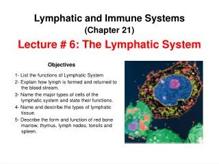
Lymphatic and Immune Systems. (Chapter 21). Lecture # 6 : The Lymphatic System. Objectives. 1- List the functions of Lymphatic System 2- Explain how lymph is formed and returned to the blood stream. 3- Name the major types of cells of the lymphatic system and state their functions.

(Chapter 21)
E N D
Presentation Transcript
Lymphatic and Immune Systems (Chapter 21) Lecture # 6: The Lymphatic System Objectives • 1- List the functions of Lymphatic System • 2- Explain how lymph is formed and returned to the blood stream. • 3- Name the major types of cells of the lymphatic system and state their functions. • 4- Name and describe the types of lymphatic tissue. • 5- Describe the form and function of red bone marrow, thymus, lymph nodes, tonsils and spleen.
Immune System It is not an organ system, but a population of cells that inhabit all of our organs and defend the body from agents of disease. Immune cells are specially concentrated in a true organ system, the Lymphatic System. Lymphatic System It is a true organ system composed of a network of vessels that penetrate nearly every tissue of the body, and a collection of tissues and organs that produce immune cells. The Lymphatic System
The Lymphatic System 1- Fluid recovery Functions of the Lymphatic System 2- Immunity 1- Fluid recovery 3- Lipid absorption • Fluid continually filters from the blood capillaries into the tissue spaces. But the blood capillaries reabsorb only 85%. 15% (2 – 4 L/day) of the water and about half of the plasma proteins are not absorbed by capillaries. • One function of the lymphatic system is to reabsorb this excess and to return it to the blood. 7 mm Hg 13 mm Hg
2- Immunity The region of overlap of endothelial cells acts as a one-way valve, permitting the entry of fluid and solutes (even those as large proteins), as well as viruses, bacteria, and cell debris, but preventing their return to the interstitial space. On its way to the blood stream, the fluid passes through lymph nodes, where immune cells stand guard against foreign matter. When they detect anything potentially harmful, they activate a protective immune response. Lymph Node Structure
3- Lipid absorption • Special lymphatic vessels called lacteals in small intestine absorb dietary lipids that are not absorbed by the blood capillaries.
The Lymphatic System Lymphatic Vessels Lymph Lymphoid Tissues Lymphoid Organs It is the recovered fluid. Lymph is usually a clear colorless fluid, similar to blood plasma but low in protein. - Lymphatic capillaries Aggregates of lympho-cytes in the connective tissue of mucous membrane and various organs. They have well –defined anatomical sites and at least partial connective tissue capsules. - Lymphatic collecting vessels 1- Diffuse lymphatic tissue 1- Red bone marrow - Lymphatic trunks 2- Thymus Mucosa-associated lymphatic tissue (MALT) 3- Lymph nodes - Collecting ducts 2- Lymphatic nodules (follicles) 4- Tonsils 5- Spleen Peyer patches in the distal portion of the small intestine
Lymph and Lymphatic Vessels Lymph: • It is the extracellular fluid drawn into lymphatic capillaries. • It is a clear, colorless fluid, similar to plasma, but contains much less protein. After a meal, lymph draining from the small intestine has a milky appearance because of its lipid contains. Lymph leaving the lymph nodes contains a large number of lymphocytes (indeed, this is the main supply of lymphocytes to the blood stream). Lymph may also contain macrophages, hormones, bacteria, viruses, cellular debris, or even cancer cells.
Lymphatic Vessels 1- Lymphatic capillaries (terminal lymphatics) • They penetrate nearly every tissue of the body, but are absent from central nervous system, cartilage, cornea, bone and bone marrow. Anchoring filaments • They are sacs of thin endothelial cells that loosely overlap each other closed at one end. Tissuefluid Endothelium of lymphatic capillary • The cells are attached to surrounding tissue by protein filaments. • The gaps between cells are large enough to allow bacteria and cells entrance to lymphatic capillary. • The endothelium creates valve-like flaps that open when interstitial fluid pressure is high, and close when it is low.
The lymphatic capillaries converge to form collecting vessels. Lymphatic system Cardiovascular system The collecting vessels travel alongside veins and arteries, and at irregular intervals they empty into lymph nodes. In the lymph nodes, bacteria are phagocytized and immune cells monitor the fluid for foreign antigens. Subclavian vein Pulmonary circuit Collecting ducts (2) Lymphatic trunks (6) The collecting vessels converge to form lymphatic trunks (6). Superior vena cava Collecting vessels There are six lymphatic trunks, whose names indicates their locations and part of the body they drain. Collecting vessels 1- Jugular trunks, 2- Subclavian trunks, 3- Bronchomediastinal trunks, 4-Inter-costal trunks, 5- Intestinal trunk, and 6- Lumbar trunks. Blood flow Lymphatic capillaries The lymphatic trunks converge to form collecting ducts: Lymphatic capillaries 1- Right lymphatic duct Systemic circuit 2- Thoracic duct
Lymph Lymph flows forward through open valves Closed valves prevent backflow
Right jugular trunk Left jugular trunk Right subclavian trunk Left subclavian trunk Right lymphatic duct Left subclavian vein Right subclavian vein Left broncho- mediastinal trunk Right broncho- mediastinal trunk Thoracic duct Cisterna chyli Intestinal trunk Right lumbar trunk Left lumbar trunk
The right lymphatic duct receives lymph from right arm, right side of head and thorax; empties into right subclavian vein. • The thoracic duct (larger and longer) receives lymph from below diaphragm, left arm, left side of head, neck, and thorax; empties into left subclavian vein.
Lymphatic Cells 1- Natural killer (NK) cells 4- Macrophages Lymphocytes 2- T lymphocytes (T cells) 5- Dendritic cells 3- B lymphocytes (B cells) 6- Reticular cells 1-Natural killer (NK) cells • They are large lymphocytes that attack and destroy bacteria, transplanted tissue, and host cells infected with viruses or that have turned cancerous. They are responsible for immune surveillance.
Red bone marrow 2- T lymphocytes (T cells) T cells are born in the red bone marrow. They are released into the blood as still-undifferentiated stem cells. T stem cell Thymus The thymus is the “school” where they mature into fully functional T lymphocytes. T lymphocyte 3- B lymphocytes (B cells) B stem cell B cells are also born in the red bone marrow, but they remain there until they differentiate in functional B lymphocytes. B lymphocyte
4- Macrophages • They are very large, avidly phagocytic cells of the connective tissue. They develop from monocytes that have emigrated from the blood stream. • They phagocytize tissue debris, dead neutrophils, bacteria, and other foreign matter. • They process foreign matter and display antigenic fragments to certain T cells, thus alerting the immune system to the presence of the enemy (they are antigen presenting cells (APCs).
5- Dendritic cells • They alert immune system to pathogens that have breached their surface. • They are branched, mobile antigen presenting cells (APCs) found in epidermis, mucous membranes, and lymphatic organs. In the skin they are called Langerhans’ cells. They engulf foreign matter by receptor mediated endocytosis rather than phagocytosis. 6- Reticular cells • They are branched stationary epithelial cells that contribute to the stroma of a lymphatic organ. They act as APCs in the thymus.
Lymphatic Tissues 1-Diffuse lymphatic tissue Lymphatic (lymphoid) tissues 2-Lymphatic nodules (follicles) They are aggregations of lymphocytes in the connective tissues of mucous membranes and various organs. They have no fibrous capsule surrounding them. 1- Diffuse lymphatic tissue It is the simplest form in which lymphocytes are scattered, rather than densely clustered. It is prevalent in the mucosa of body passages open to the exterior respiratory, digestive, urinary, and reproductive tracts, where it is called mucosa-associated lymphatic tissue (MALT) 2- Lymphatic nodules (follicles) They are dense masses of lymphocytes and macrophages that congregate in some places. - Appendix • - Peyer patches: They are dense clusters in the ileum, the distal portion of the small intestine.
Lymphatic Organs Lymphatic organs have well-defined anatomical sites and have connective tissue capsule that separates the lymphatic tissue from neighboring tissues. Primary lymphatic organs • Red bone marrow • They are the sites where T and B cells become immunocompetent (able to recognize and respond to antigens). Lymphatic organs Thymus Lymph nodes Secondary lymphatic organs They are populated with lymphocytes after these cells are mature (immunocompetent). Tonsils Spleen Immunocompetency: It is the ability to recognize and respond to antigens. Antigen: It is any molecule that triggers an immune response.
Red Bone Marrow • It is a soft, loosely organized, highly vascular material separated from osseous tissue by endosteum of bone. • As blood cells mature, they push their way through the reticular and endothelial cells to enter the sinusoids and flow away in the blood stream. Functions: 1- It is involved in hemopoiesis (blood formation). Erythrocytes (RBCs), leukocytes (WBCs), and platelets are produced in the red bone marrow. 2- It is an important supplier of lymphocytes to the immune system.
Thymus Thyroid The thymus is a member of the endocrine, lymphatic, and immune systems. Trachea Thymus • It is a bilobed organ located in superior mediastinum between the sternum and aortic arch. • The fibrous capsule gives off trabeculae (septa) that divide the gland into several lobules. • The lobules have cortex and medulla populated by T lymphocytes. Functions: • 1- It houses developing lymphocytes. • 2- The reticular epithelial cells produce signaling molecules (thymosin, thymopoietin, thymulin, interleukins, and interferon) that promote the development and action of T lymphocytes.
Lymph Nodes They are the most numerous lymphatic organs (about 450 in typical young adult). They are elongated, bean shaped structures with an indentation called the hilum. They are enclosed with a fibrous capsule with trabeculae that divide the interior into compartments. The parenchyma is divided into cortex and medulla. • The cortex contains germinal centers where B cells multiply and differentiate into plasma cells. Several afferent lymphatic vessels lead into the node along its convex surface. • Lymph leaves the node through one to three efferent lymphatic vessels that leave the hilum.
Lymph nodes are wide-spread but specially concentrated in some locations. Cervical lymph nodes R. lymphatic duct Thoracic duct Axillary lymph node Thymus Thoracic duct Cisterna chyli Spleen Abdominal, intestinal, and mesenteric lymph nodes Functions: Inguinal lymph nodes • 1- They cleanse the lymph. • 2- They act as a site of T and B cell activation. Popliteal lymph nodes
Tonsils • They are patches of lymphatic tissue located at the entrance to the pharynx. Each tonsil is covered with epithelium and have deep pits (tonsillar crypts) lined with lymphatic nodules. Pharyngeal tonsil or adenoids (1) Functions: Palatine tonsil (2) • They guard against ingested or inhaled pathogens. Lingual tonsil (numerous)
Spleen It is the body’s largest lymphatic organ. Functions: • 1- Blood production in fetus. Sinuses filled with erythrocytes • 2- It helps to stabilize blood volume by transferring excess plasma from the blood to the lymph. • 3- It is an “erythrocyte graveyard” ( RBC disposal). • 4- Lymphocytes and macrophages of white pulp monitors blood for foreign antigens, much like the lymph nodes do the lymph. Lymphocytes, macrophages and small branches of splenic artery.