Download
1 / 83
1.18k likes | 2.51k Vues
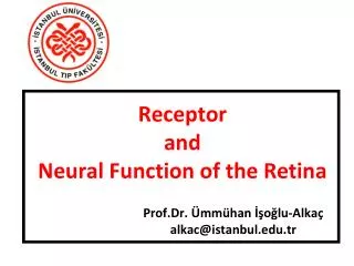
Receptor and Neural Function of the Retina. Prof.Dr. Ümmühan İşoğlu-Alkaç alkac@istanbul.edu.tr. Retina. The retina is the light-sensitive portion of the eye that contains 1) the cones , which are responsible for color vision, and

E N D
Receptor andNeural Function of the Retina Prof.Dr. Ümmühan İşoğlu-Alkaç alkac@istanbul.edu.tr
Retina • The retina is the light-sensitive portion of the eyethat contains 1) the cones, which are responsiblefor color vision, and 2) the rods, which are mainlyresponsible for black and white vision and vision in the dark.
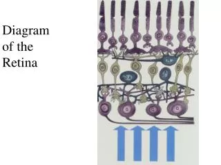
Layers of the Retina layers from the outside to the inside as follows: 1) pigmented layer, 2) layer of rods and cones projecting to the pigment, 3) outer nuclear layer containingthe cell bodies of the rods and cones, 4) outer plexiform layer, 5) Innernuclear layer, 6) Inner plexiform layer, 7) ganglionic layer, 8) layer of optic nervefibers, and 9) inner limiting membrane.
Foveal Region of the Retina and Its Importance in Acute Vision. • The fovea is a minute areain the center of the retina, it is especially capable of acute and detailed vision (1mm2). • Thecentral fovea (0.3 mm2), is composed almost entirely of cones;these cones have a special structure that aids their detection of detail in the visualimage. • In the foveal region, the blood vessels, ganglion cells, inner nuclear layer of cells,and plexiform layers are all displaced to one side rather than resting directly on topof the cones. This allows light to pass unimpeded to the cones.
Photomicrograph of the macula and of the fovea in its center. Note that the inner layers of the retina are pulled to the side to decrease interference with light transmission. (From Fawcett DW: Bloom and Fawcett: A Textbook of Histology, 11th ed. Philadelphia: WB Saunders, 1986; courtesy H. Mizoguchi.)
Rod-Cone the major functional segments of either arod or a cone: • the outer segment, • the inner segment, • the nucleus, and • the synaptic body.
The light-sensitive photochemical is found inthe outer segment. • In the case of the rods, this is rhodopsin; in the cones, it is one of three “color” photochemicals, usually called simply color pigments,that function almost exactly the same as rhodopsinexcept for differences in spectral sensitivity.
The inner segment of the rod or cone contains theusual cytoplasm with cytoplasmic organelles. • Particularlyimportant are the mitochondria; as explained later,these mitochondria play the important role of providingenergy for function of the photoreceptors.
The synaptic body is the portion of the rod or conethat connects with subsequent neuronal cells, thehorizontal and bipolar cells, that represent the nextstages in the vision chain.
Schematic drawing of the functional parts of the rods and cones.
Membranous structures of the outer segments of a rod (left) anda cone (right) (Courtesy Dr. Richard Young.)
Pigment Layer of the Retina • The black pigment melanin inthe pigment layer prevents light reflection throughoutthe globe of the eyeball; this is extremely important forclear vision. This pigment performs the same functionin the eye as the black coloring inside the bellows of a camera.Without it, light rays would be reflected in alldirections within the eyeball and would cause diffuse lighting of the retina rather than the normal contrastbetween dark and light spots required for formation ofpreciseimages.
The importance of melanin in the pigment layer iswell illustrated by its absence in albinos, people who arehereditarily lacking in melanin pigment in all parts of their bodies. The pigment layer also stores large quantities ofvitamin A. • This vitamin A is exchanged back and forththrough the cell membranes of the outer segments ofthe rods and cones, which themselves are embedded inthe
Blood Supply of the Retina Central Retinal Artery and theChoroid. • The nutrient blood supply for the internallayers of the retina is derived from the central retinalartery, which enters the eyeball through the center ofthe optic nerve and then divides to supply the entireinside retinal surface. Thus, the inner layers of the retinahave their own blood supply independent of the other structures of the eye.
However, the outermost layer of the retina is adherentto the choroid, which is also a highly vascular tissuelying between the retina and the sclera. • The outer layersof the retina, especially the outer segments of the rodsand cones, depend mainly on diffusion from the choroidblood vessels for their nutrition, especially for their oxygen
Retinal Detachment • The neural retina occasionallydetaches from the pigment epithelium.
Photochemistry of Vision • Both rods and cones contain chemicals that decomposeon exposure to light and, in the process, excite thenerve fibers leading from the eye. • The light-sensitivechemical in the rods is called rhodopsin; the lightsensitivechemicals in the cones, called cone pigmentsor color pigments, have compositions only slightly different from that of rhodopsin.
Rhodopsin-retinal visual cycle in the rod, showing decompositionof rhodopsin during exposure to light and subsequent slow reformationof rhodopsin by the chemical processes.
Vitamin A • Vitamin A is present both in the cytoplasm of the and in the pigment layer of theretina.Therefore, vitamin A is normally always available to form newretinal when needed. Conversely, when there isexcessretinal in the retina, it is converted back into vitaminA, thus reducing the amount of light-sensitive pigmentin the retina.
Night Blindness • Night blindness occurs in any personwith severe vitamin A deficiency.
Dark current origins. ATP adenosine triphosphate; cGMP cyclic guanosine monophosphate; GTP guanosine triphosphate.
Phototransduction under low light conditions. ATP adenosine triphosphate; cGMP cyclic guanosine monophosphate.
Photosensory transduction pathway and mechanisms for limiting and terminating signaling. cGMP cyclic guanosine monophosphate
Theoretical basis for generation of a “hyperpolarization receptorpotential” caused by rhodopsin decomposition, which decreasesthe flow of positively charged sodium ions into the outer segmentof the rod.
Automatic Regulation of Retinal Sensitivity—Light and Dark Adaptation • If a person has been in brightlight for hours, large portions of thephotochemicals inboth the rods and the cones will have been reduced toretinal and opsins. Furthermore, much of the retinal ofboth the rods and the cones will have been convertedinto vitamin A.
Because of these two effects, theconcentrationsof the photosensitive chemicals remainingin the rods and cones are considerably reduced, andthesensitivity of the eye to light is correspondinglyreduced. This is called light adaptation.
Conversely, if a person remains in darkness for along time, • the retinal and opsins in the rods and conesare converted back into the light-sensitive pigments. • Furthermore, vitamin A is converted back into retinalto give still more light-sensitive pigments, the finallimit being determined by the amount of opsins in therods and cones to combine with the retinal. This is called dark adaptation.
Dark adaptation, demonstrating the relation of cone adaptation to rod adaptation.
Other Mechanisms of Light and Dark Adaptation. • 1-In additionto adaptation caused by changes in concentrations ofrhodopsin or color photochemicals, the eye has twoother mechanisms for light and dark adaptation. • 2- Thefirst of these is a change in pupillary size. This can cause adaptation of approximately30-fold within a fraction of a second, because ofchanges in the amount of light allowed through the pupillary opening.
3-The other mechanism is neural adaptation, involvingthe neurons in the successive stages of the visual chainin the retina itself and in the brain. That is, when lightintensity first increases, the signals transmitted by thebipolar cells, horizontal cells, amacrine cells, and ganglioncells are all intense. However, most of these signalsdecrease rapidly at different stages of transmission inthe neural circuit.
Although the degree of adaptation isonly a fewfold rather than the many thousandfold thatoccurs during adaptation of the photochemical system, neural adaptation occurs in a fraction of a second, incontrast to the many minutes to hours required for full adaptation by the photochemicals.
Tricolor Mechanism of Color Detection All theories of color vision are based on the wellknownobservation that the human eye can detectalmost allgradations of colors when only red, green,and blue monochromatic lights are appropriately mixed in different combinations.
Spectral Sensitivities of the Three Types of Cones On thebasis of color vision tests, the spectral sensitivities ofthe three types of cones in humans have proved to beessentially the same as the light absorption curves forthe three types of pigment found in the cones.They can explain most of the phenomena of color vision.
Cones; • Blue (445 nm) • Green (535 nm) • Red (570 nm) sensitive pigment
Demonstration of the degree of stimulation of the different colorsensitivecones by monochromatic lights of four colors: blue, green, yellow, and orange.
Color Vision; Interpretation of Color in the Nervous System Referringone can see that an orangemonochromaticlight with a wavelength of 580nanometers stimulates the red cones to a stimulusvalue of about 99; it stimulates the green conesto a stimulus value of about 42, but the blue cones notat all. Orange: red 99: green 42: blue 0. The nervoussystem interprets this set of ratios as the sensation of orange. • Blue: 0:0:97; Yellow:83:83:0; Green:31:67:36
Farklı renklere duyarlı konilerin mavi, yeşil, sarı ve turuncu monokromatik ışıklarla uyarılma dereceleri.
Perception of White Light About equal stimulation ofall the red, green, and blue cones gives one the sensation of seeing white.
Red-Green Color Blindness • When a single group of colorreceptivecones is missing from the eye, the person isunable to distinguish some colors from others. A person with loss of red cones is called a protanope;the overall visual spectrum is noticeably shortened atthe long wavelength end because of a lack of the redcones. A color-blind person who lacks green cones iscalled a deuteranope; this person has a perfectly normalvisual spectral width because red cones are available todetect the long wavelength red color.
Red-green color blindness is a genetic disorder thatoccurs almost exclusively in males. That is, genes in thefemale X chromosome code for the respective cones. • Becausethe male has only one X chromosome, a missing genecan lead to color blindness. • colorblindness is passed from mother to son, and the motheris said to be a color blindness carrier; this is true in about 8 % of all women.
Ishihara Test. The normal personreads “74,” the red-green color-blind person reads “21.”The red-blind person (protanope) reads“2,” but the green-blind person (deuteranope) reads “4.” Thenormal person reads “42.” (Reproduced from Ishihara’s Testsfor Colour Blindness.Tokyo: Kanehara&Co.)
Neural Function of the Retina The different neuronal cell types are as follows: 1. The photoreceptors themselves—the rods andcones—which transmit signals to the outerplexiform layer, where they synapse with bipolar cells and horizontal cells 2. The horizontal cells, which transmit signalshorizontally in the outer plexiform layer from therods and cones to bipolar cells 3. The bipolar cells, which transmit signals verticallyfrom the rods, cones, and horizontal cells to theinner plexiform layer, where they synapse withganglion cells and amacrine cells