Chapter 34 Organization and Control of Neural Function
360 likes | 630 Vues
Essentials of Pathophysiology. Chapter 34 Organization and Control of Neural Function. The dorsal horn cell columns contain the afferent (sensory) neurons and the ventral horn cell columns contain the efferent neurons.

Chapter 34 Organization and Control of Neural Function
E N D
Presentation Transcript
Essentials of Pathophysiology Chapter 34Organization and Control ofNeural Function
The dorsal horn cell columns contain the afferent (sensory) neurons and the ventral horn cell columns contain the efferent neurons. • The brain is divided into three regions: the hindbrain, the midbrain, and the forebrain. • The parasympathetic nervous system functions in maintaining vital functions and responding when there is a critical threat to the integrity of the individual—the “fight-or-flight” response. • The blood-brain barrier and the cerebrospinal fluid–brain barrier protect the brain from substances in the blood that would disrupt brain function. • Cerebrospinal fluid helps maintain a constant ionic environment that serves as a medium for diffusion of nutrients, electrolytes, and metabolic end products into the extracellular fluid surrounding central nervous system neurons and glia. Pre lecture quiz T T F T T
______________ are the functioning cells of the nervous system. • The _________________ nervous system contains two divisions: sympathetic and parasympathetic. • Inside the skull and vertebral column, the brain and spinal cord are loosely suspended and protected by several connective tissue sheaths called the _________________. • Neurons communicate with each other through structures known as ________________, of which there are two types: electrical and chemical. • The main ___________________ for the autonomic nervous system are acetylcholine and the catecholamines, epinephrine and norepinephrine, which control neural function by selectively causing excitation or inhibition of action potentials. Pre lecture quiz • Autonomic • meninges • Neurons • neurotransmitters • synapses
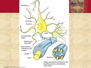
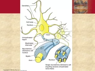
Dendrites receive stimuli Stimuli pass down axons Schwann cells contain myelin (“white matter”) Help increase speed of impulse transmission Neurons
Peripheral nervous tissue • Schwann cells: wrap a layer of myelin around axons • Satellite cells: separate nervous cells from supporting tissue • Central nervous tissue • Oligodendroglia: myelinating cells • Astroglia: regulate ion content in intercellular fluid • Microglia: phagocytes • Ependymal cells: line the neural tube cavity Supporting Cells
Microglial cell Neuron Oligodendritic cell Ependymal cell Astrocyte Identify E C B D What is the function of each? A
Tell whether the following statement is true or false. All neurons are myelinated. Question
False Rationale:The myelin sheath increases the speed of impulse transmission (the impulse can skip over the myelinated/insulated parts of the neuron), but speed is not important everywhere (like the digestive tract). If every neuron was myelinated, neurons would take up a lot more space, too. Answer
Stimulus opens Na+ gates At threshold, more Na+ gates open Na+ enters cell: depolarization K+ gates open K+ diffuses out: repolarization The Basics of Cell Firing Action potential Threshold potential Resting membrane potential Stimulus
What is happening at stages 1–4? What will result if you block stage 2? Stage 3? Stage 4? Synaptic Transmission
Neurotransmitters • Amino acids • Peptides • Monoamines • Neuromodulators • Attach to receptors and change their response to neurotransmitters • Neurotrophic factors • Neuron survival and to develop connections between neurons Neuron Secretions
Begins as a hollow tube • First segments of the tube become the brain • Forebrain • Midbrain • Hindbrain General Organization of the Nervous System
Dorsal • Afferent • Sensory • Ventral • Efferent • Motor Organization of the Spinal Cord
A woman developed polyneuropathy. • Her spinal nerves were damaged • She lost the ability to tell where her body was positioned • She has to look every time she takes a step, to tell where she is moving her feet to Question: • What parts of her spinal nerves were damaged? Scenario
What problems would you expect in someone who suffered ischemia to: • Area A • Area B • Area C Cell Columns of the Spinal Cord A B C
If you place your hand on a hot surface, which ganglion carries the impulse to the spinal cord? • Ventral • Dorsal • Interneuron • Association neuron Question
Dorsal Rationale:Afferent neurons carry sensory impulses to the spinal cord through the dorsal root ganglion; efferent neurons carry motor responses through the ventral root ganglion to effector cells in the tissue. Answer
Archi layer • Connects neighboring segments • Contains neurons reticular activating system • Paleo layer • Fibers reach to the brain stem • Neo layer • Pathways for bladder control and fine motor skills • Develop by fifth year of life Layers of the White Matter
Cerebrum Thalamus Hypothalamus Cerebral peduncles Cerebral aqueduct Colliculi Cerebellum Pons Medulla oblongata Brain Regions
Medulla oblongata, cerebellum, and pons Reflex centers for heart and respiration rates, coughing, swallowing, vomiting, etc. Gives rise to cranial nerves V–XII controlling viscera, hearing, facial, and mouth/throat functions Cerebellum allows fine motor coordination Functions of the Hindbrain
Cerebral peduncles carry nerve fibers from the cerebrum to the hindbrain Cerebral aqueduct lets cerebrospinal fluid drain from the fourth ventricle inside the cerebrum Superior colliculi control reflex eye movements Inferior colliculi control reflex reactions to sound Gives rise to cranial nerves III and IV, controlling eye movement Functions of the Midbrain
Thalamus: “switchboard” or relay station for impulses going to and coming from the cerebrum Hypothalamus: homeostatic control Cerebrum Gives rise to cranial nerves I and II, for smell and sight Functions of the Forebrain
Frontal lobe: motor, anticipation Parietal lobe: somatosensory Temporal lobe: hearing, memory Occipital lobe: vision Limbic system: emotional Cerebrum
Which part of the brain maintains vital functions like breathing, heart rate, and digestion? • Forebrain • Midbrain • Hindbrain • Cerebellum Question
Hindbrain Rationale:Also known as the brain stem, this is the vasomotor center that controls cardiopulmonary function and digestion. Answer
Has two layers Inner layer bends over to form a fold (falxcerebri) that separates the cerebral hemispheres It forms a second fold (tentorium) that holds the cerebrum up off the cerebellum Dura Mater
Between the layers of the dura, at the base of each fold, venous blood drains out of the brain in a sinus Bridging veins carry blood from the brain across the inner layer of the dura mater to the sinus The sinus also collects cerebrospinal fluid Dura Mater (cont.)
Lies just beneath the dura mater Waterproof Cerebrospinal fluid (CSF) lies under the arachnoid to cushion the brain Extensions of the arachnoid (villi) poke through the inner layer of the dura mater into the sinuses, to let CSF drain into the sinuses Arachnoid
Lies right on the surface of the brain Holds the cerebral arteries in place Pia Mater
Epidural space: meningeal arteries • Dura mater • Subdural space: bridging veins • Arachnoid • Subarachnoid space: cerebral arteries, cerebrospinal fluid • Pia mater Meninges and Meningeal Spaces
Leaks out of capillaries inside the brain’s hollow ventricles • Composition controlled by the blood-brain barrier • Passes out an opening below the cerebellum • Circulates around the brain and spinal cord in the subarachnoid space • Passes through arachnoidvilli into blood in the dural sinuses and is returned to the heart Cerebrospinal Fluid
Sympathetic • Catecholamines • Epinephrine, norepinephrine, dopamine • Attach to adrenergic receptors • Parasympathetic • Acetylcholine • Attaches to cholinergic receptors Autonomic Nervous System
Synthesized in the sympathetic system • Attach to adrenergic receptors • Alpha-1 receptors: constrict blood vessels • Alpha-2 receptors: negative feedback to stop neurotransmitter release • Beta-1 receptors: speed and strengthen heart • Beta-2 receptors: bronchodilation • Neurotransmitter is removed from synapse by reuptake or degraded by enzymes Adrenergic Neurotransmitters
Released from parasympathetic system and from motor neurons • Attaches to cholinergic receptors • Nicotinic receptors: excite skeletal muscle cells • Muscarinic receptors: slow heart, stimulate GI tract, vasodilate • Neurotransmitter is removed from synapse by acetylcholinesterase Cholinergic Neurotransmitter—Acetylcholine
Tell whether the following statement is true or false. The sympathetic division of the ANS is also known as fight-or-flight. Question
True Rationale:The SNS is characterized by the release of adrenaline, which results in pupil dilation, bronchodilation, and increased HR, BP, and glucose production—all the things that come in handy when you are running from something! Answer