Download
1 / 44
460 likes | 818 Vues
Ventricular Tachyarrhythmias. An Electrophysiologic Overview. Module Objectives – Ventricular Tachyarrhythmias. After completion of this module, the participant should be able to:. Identify the mechanisms for ventricular tachycardias

E N D
Ventricular Tachyarrhythmias An Electrophysiologic Overview
Module Objectives – Ventricular Tachyarrhythmias After completion of this module, the participant should be able to: • Identify the mechanisms for ventricular tachycardias • Differentiate types of ventricular tachycardias using ECG and intracardiac electrogram recordings • Discuss treatment options for ventricular tachycardias
Module Outline – Ventricular Tachyarrhythmias • Description • Characteristics • Mechanisms • Sustained vs. nonsustained • Premature ventricular contractions
Module Outline – Ventricular Tachyarrhythmias • Classification • Monomorphic • Idiopathic • Description • ECG recognition • Treatment – ablation • Bundle branch • Description • ECG recognition • Treatment –ablation
Module Outline – Ventricular Tachyarrhythmias • Classifications - continued • Ventricular flutter • ECG recognition • Ventricular fibrillation • ECG recognition • Polymorphic • Torsades de pointes • Description • ECG recognition • Treatment • Summary
Ventricular Tachycardia (VT) • Originates in the ventricles • Can be life threatening • Most patients have significant heart disease • Coronary artery disease • A previous myocardial infarction • Cardiomyopathy
Mechanisms of VT • Reentrant • Reentry circuit (fast and slow pathway) is confined to the ventricles and/or bundle branches • Automatic • Automatic focus occurs within the ventricles • Triggered activity • Early afterdepolarizations (phase 3) • Delayed afterdepolarizations (phase 4)
Reentrant • Reentrant ventricular arrhythmias • Premature ventricular complexes • Idiopathic left ventricular tachycardia • Bundle branch reentry • Ventricular tachycardia and fibrillation when associated with chronic heart disease: • Previous myocardial infarction • Cardiomyopathy
Automatic • Automatic ventricular arrhythmias • Premature ventricular complexes • Ischemic ventricular tachycardia • Ventricular tachycardia and fibrillation when associated with acute medical conditions: • Acute myocardial infarction or ischemia • Electrolyte and acid-base disturbances, hypoxemia • Increased sympathetic tone
Abnormal Acceleration of Phase 4 Automaticity Fogoros: Electrophysiologic Testing. 3rd ed. Blackwell Scientific 1999; 16.
Triggered • Triggered activity ventricular arrhythmias • Pause-dependent triggered activity • Early afterdepolarization (phase 3) • Polymorphic ventricular tachycardia • Catechol-dependent triggered activity • Late afterdepolarizations (phase 4) • Idiopathic right ventricular tachycardia
Triggered Fogoros: Electrophysiologic Testing. 3rd ed. Blackwell Scientific 1999; 158.
Sustained vs. Nonsustained • Sustained VT • Episodes last at least 30 seconds • Commonly seen in adults with prior: • Myocardial infarction • Chronic coronary artery disease • Dilated cardiomyopathy • Non-sustained VT • Episodes last at least 6 beats but < 30 seconds
Premature Ventricular Contraction • PVC • Ectopic beat in the ventricle that can occur singly or in clusters • Caused by electrical irritability • Factors influencing electrical irritability • Ischemia • Electrolyte imbalances • Drug intoxication
Classification • Ventricular Tachycardia • Monomorphic • Idiopathic VT • Bundle branch reentry tachycardia • Ventricular flutter • Ventricular fibrillation • Polymorphic • Torsades de pointes (TdP)
Monomorphic VT • Heart rate: 100 bpm or greater • Rhythm: Regular • Mechanism • Reentry • Abnormal automaticity • Triggered activity • Recognition • Broad QRS • Stable and uniform beat-to-beat appearance
ECG Recognition ECG used with permission of Dr. Brian Olshansky.
Intracardiac Recording of VT EGM used with permission of Texas Cardiac Arrhythmia, P.A.
Idiopathic Right Ventricular Tachycardia • Right ventricular idiopathic VT • Focus originates within the right ventricular outflow tract • Ventricular function is usually normal • Usually LBBB, inferior axis • Treatment options: • Pharmacologic therapy (beta blockers, verapamil) • RF ablation
ECG Recognition Kay NG. Am J Med 1996; 100: 344-356.
Case History: Idiopathic VT 39 y.o. female with no prior cardiac history • First episode • 9 hours of palpitations • In ER, found to be in wide-complex tachycardia of LBBB, inferior axis, at 205 bpm • Converted with IV lidocaine; placed on tenormin • Second episode • While on tenormin, patient had onset of palpitations at airport • In ER, converted with IV lidocaine • Patient underwent EP study
Case History: Idiopathic VT • At EP study, tachycardia focus was mapped and localized to right ventricular outflow tract • The focus was successfully ablatedusing radiofrequency energy, with no subsequent inducible or clinical VT
Endocardial Activation Mapping • Using an ablation catheter, map the area around and inside of the right ventricular outflow tract • Find the electrograms that precede the onset of the QRS complex during tachycardia • This area identifies the site of earliest activation, and possibly the “site of origin” of the arrhythmia
Pace Mapping • Pace mapping helps to localize the “site of origin” after endocardial mapping has been performed • If the heart is paced from this region, the resulting ECG should be identical to the ECG taken during tachycardia • Delivering RF energy to this site usually eliminates ventricular tachycardia
Idiopathic VT Ablation in RVOT RAO RAO
Idiopathic Left Ventricular Tachycardia • RBBB/LAFB • Involves the Purkinje network • Treatment options: • RF ablation • Pharmacologic therapy (verapamil, beta blockers)
ECG Recognition ECG used with permission of Kay NG.
Bundle Branch Reentry • Reentry circuit is confined to the left and right bundle branches • Usually LBBB, during sinus rhythm • Presents with: • Syncope • Palpitations • Sudden cardiac death • Treatment: RF ablation of right bundle
Catheter Ablation of Right Bundle Branch I II V1 RA Current Voltage Courtesy of Dr. Warren Jackman
Ventricular Flutter • Heart rate: 300 bpm • Rhythm: Regular and uniform • Mechanism: Reentry • Recognition: • No isoelectric interval • No visible T wave • Degenerates to ventricular fibrillation • Treatment: Cardioversion
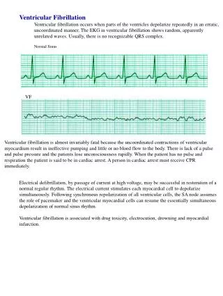
Ventricular Fibrillation • Heart rate: Chaotic, random and asynchronous • Rhythm: Irregular • Mechanism: Multiple wavelets of reentry • Recognition: • No discrete QRS complexes • Treatment: • Defibrillation
P waves and QRS complexes not present Heart rhythm highly irregular Heart rate not defined ECG Recognition
Polymorphic VT • Heart rate: Variable • Rhythm: Irregular • Mechanism: • Reentry • Triggered activity • Recognition: • Wide QRS with phasic variation • Torsades de pointes
ECG Recognition EGM used with permission of Texas Cardiac Arrhythmia, P.A.
Torsades de Pointes (TdP) • Heart rate: 200 - 250 bpm • Rhythm: Irregular • Recognition: • Long QT interval • Wide QRS • Continuously changing QRS morphology
Mechanism • Events leading to TdP are: • Hypokalemia • Prolongation of the action potential duration • Early afterdepolarizations • Critically slow conduction that contributes to reentry
QRS morphology continuously changes Complexes alternates from positive to negative ECG Recognition
Possible Causes • Drugs that lengthen the QT: • Quinidine • Procainamide • Sotalol • Ibutilide • Physical • Ischemia • Electrolyte abnormalities
Treatment • Pharmacologic therapy: • Potassium • Magnesium • Isoproterenol • Possibly class Ib drugs (lidocaine) to decrease refractoriness/shorten length of action potential • Overdrive ventricular pacing • Cardioversion
Summary • VT ablation is not an FDA-approved indication • RF catheter ablation can be a useful technique in patients with ventricular tachycardia • Success largely depends on the etiology of the arrhythmia • Unstable sustained VT, polymorphic VT and ventricular fibrillation are not ablatable • Improved catheters and imaging techniques may change this in the future